
Vestibular
The vestibular system is a critical sensory pathway for the brain’s representation of space and motor control, providing a sensory readout of the acceleration, velocity, position of the head via an elaborate system of sensory organs located internal to the ears. These signals are integrated with somatosensory, proprioceptive, and visual signals in the cerebellum to provide an accurate and effective representation of the current head position, which is essential for motor control. Motor actions are driven largely by sensory inputs arising from the sensory systems located in the head (eyes, ears, nose) and thus knowing the current orientation and motion parameters of the head is essential in being able to generate properly calibrated motor actions.
Furthermore, our ability to navigate through large spaces depends critically on a continuously updated representation of the orientation of our body within the larger spatial environment, known as head direction representations. These are computed from inputs originating deep in the vestibular nuclei.
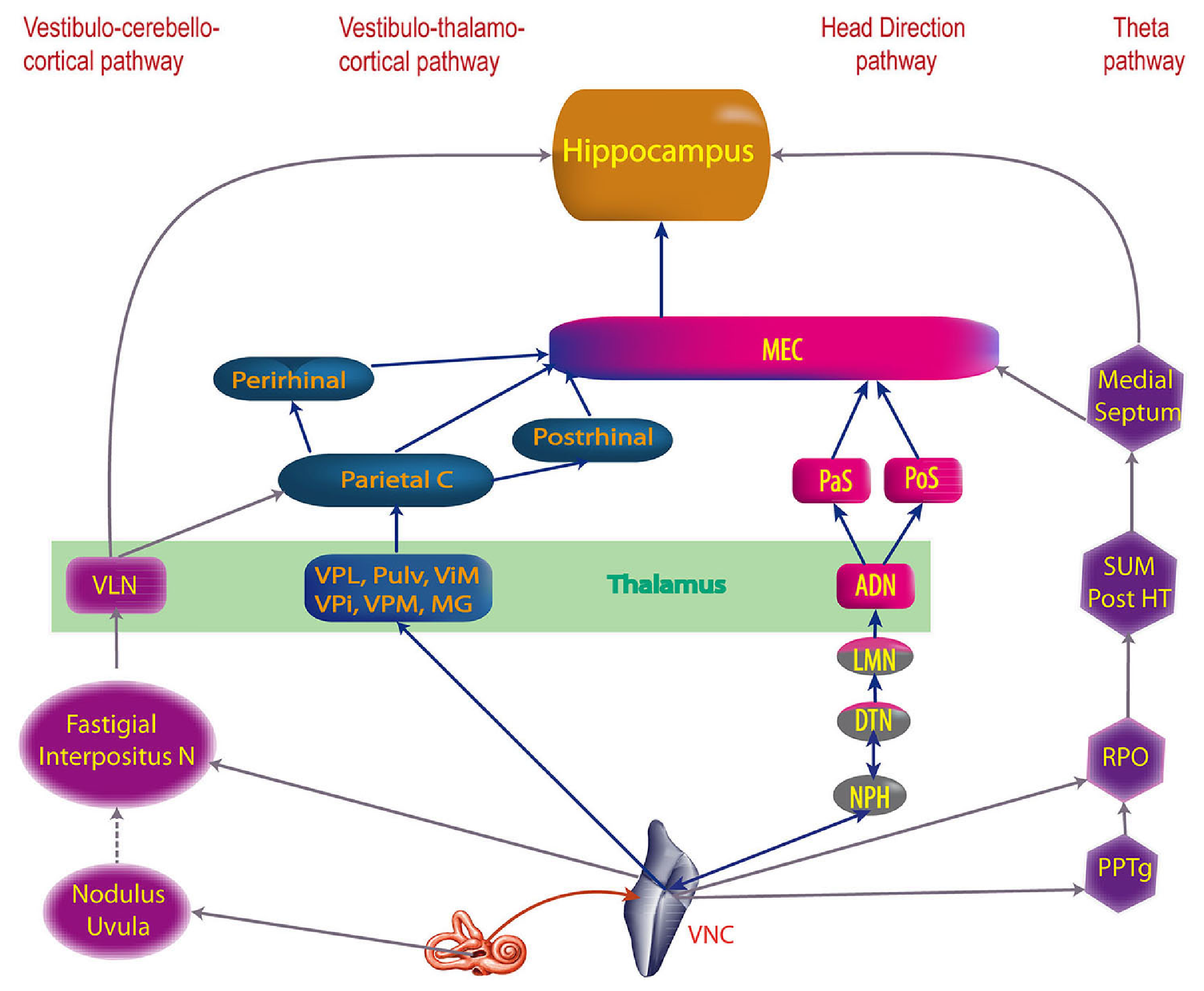
Figure 1: Overview of the vestibular system pathways, originating in the vestibular nuclear complex (VNC), which is strongly bidirectionally interconnected with the cerebellum (fastigial and interpositus cerebellar nuclei). The parietal cortex somatosensory, spatial and motor areas are activated via the ventral lateral thalamus (VLN) and areas of the ventral posterior (VP; primary somatosensory), medial geniculate (MG; primary auditory), and other thalamic pathways providing direct vestibular nucleus signals. The head direction system originates in the nucleus prepositus hypoglossi (NPH) to dorsal tegmental nucleus of Gudden (DTN), which encode head and body velocity and position signals, that then drive a ring attractor computation in the lateral mammillary nuclei (LMN) where head direction cells originate, and then project to the anteriodorsal thalamus (ADN), projecting from there into the subiclum (PaS, PoS). There are also vestibular inputs to the pedunculopontine tegmental nucleus (PPTg) which is involved with the supermammillary nucleus (SUM) and posterior hypothalamus (Post HT) that drive theta activity in the medial septum. From Hitier et al., 2014.
Figure 1 (from Hitier et al., 2014) provides an overview of the vestibular system and its projections into the rest of the brain, organized into four primary tracts.
• Cerebellum: the most evolutionarily ancient parts of the cerebellum are strongly bidirectionally interconnected with the vestibular system, via the fastigial and interpositus (interposed) deep cerebellar nuclei (DCN). The central role of vestibular signals for motor processing is reflected in the massive interconnectivity of these systems.
• Direct to neocortex: the most direct projection into the neocortex, specifically the somatosensory, spatial, and motor systems in the parietal lobe, goes via a range of ventral thalamic nuclei, including areas of the ventral posterior (VP) and medial geniculate (MG, medial portion) nuclei. The VP is primary somatosensory thalamus and MG is primary auditory thalamus, both of which have strong subcortical driver inputs conveying sensory information to the cortex. Sensibly, the vestibular system contributes to both of these.
• Head direction: there are several midbrain nuclei involved in computing the head direction signal from vestibular (and motor) sources, with the dorsal tegmental nucleus (DTN) (of Gudden) being the convergent area that has many different representations of head and body motion signals, which are then integrated in the lateral mammillary nucleus (LMN) where head direction cells first emerge. The LMN then provides strong driver inputs to the AD (anterodorsal) thalamus which sends these signals to the subiculum areas that are part of the broader hippocampal system. These head direction cells and more integrative spatial representations in the subiculum provide the core spatial anchors to the memory functions of the hippocampus.
• Theta rhythm: the vestibular system also provides inputs to the supermammillary (SUM) and medial septum theta rhythm networks, which are also driven by rhythmic activity from the ventral tegmental nucleus (VTN) (of Gudden).
Vestibular transduction
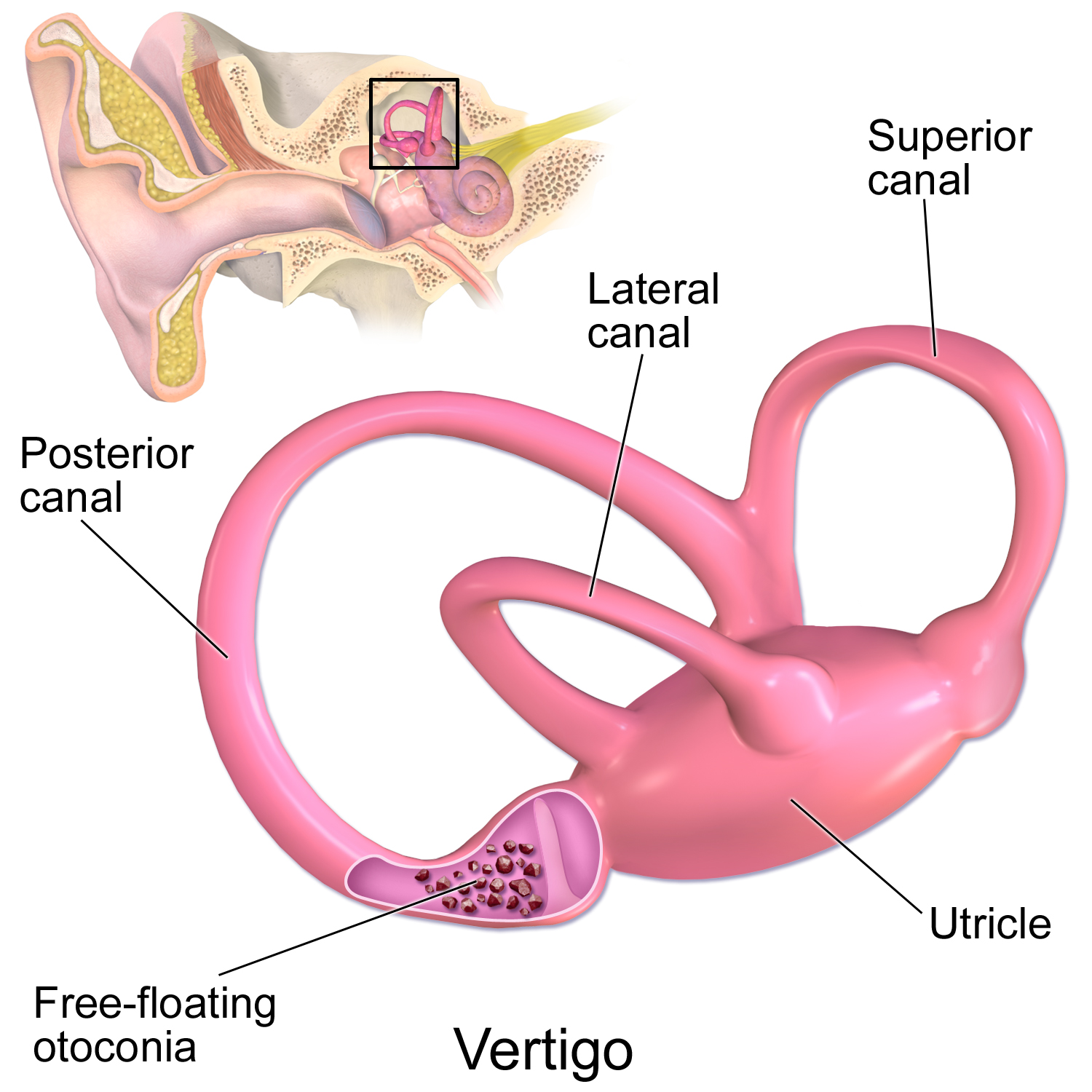
Figure 2: The vestibular organ that transduces rotational and linear motion signals into neural firing signals. The semicircular canals are sensitive to rotational motion on 3 different axes, while the otolith organs are sensitive to linear motion. Figure from BruceBlaus on Wikimedia Commons (CC-SA-4.0).
The transduction of head movements into neural firing signals occurs via the vestibular organ (Figure 2), where the semicircular canals are sensitive to rotational motion in 3 different axes, while the otolith organs are sensitive to linear accelerations (utricle for horizontal and saccule for vertical).
Vestibular nuclei (VNC)
The vestibular nuclei in the VNC can be subdivided into the descending, lateral, medial and superior nuclei (DVN, LVN, MVN, SVN), along with the parasolitary nucleus (Psol), which differ more in their projection targets than in their afferent inputs from primary vestibular signals (Barmack, 2023). These primary afferent inputs are exclusively ipsilateral (e.g., left ear to left hemisphere), but the output pathways from the nucleus neurons can project ipsilaterally or contralaterally.
There is extensive cross-hemisphere connectivity between the different nuclei, which in general establishes an opponent relationship between these neurons: any excitation of one side will lead to a proportional inhibition on the other side. This is specific to neurons that process horizontally oriented signals (semicircular canals and utricle otoliths).
In addition to primary vestibular inputs, these VNC neurons receive visual motion signals from the accessory optic system (AOS) and neck proprioceptive signals.
The different subnuclei within the VNC are not strongly functionally organized, with neurons in multiple different subnuclei projecting to the various target areas, including the cerebellar vermis and paraflocculus, extraocular muscles for eye control (e.g., for the vestibulo-ocular reflex), various areas within the MDJ (e.g., nucleus interstitialis of Cajal, subparafasicular complex, nucleus of Darkschewitsch), and the thalamus (VP).