
Synaptic plasticity
Synaptic plasticity is the well-established biological phenomenon that the strength of synaptic connections (i.e., weights) change in strength, which is known to be the primary basis for learning in the brain, consistent with the foundational hypothesis of abstract neural network models: knowledge is encoded in synaptic weights.
• LTP = long-term potentiation is the name for the phenomenon of persistent increases in synaptic strength as a result of neural activity, as initially established by Bliss & Lomo (1973).
• LTD = long-term depression refers to decreases in synaptic strength, which took a while longer to establish (Bear & Malenka, 1994).
Uncovering the biochemical basis for these phenomena has been one of the most successful collective research projects in neuroscience, resulting in a detailed understanding of many of the pathways and processes involved, which unfold over many different time scales as we briefly review here.
In an ideal world, the result of this effort would be a complete bottom-up grounding of the functional nature of learning in the neocortex: we should be able derive the learning algorithm at an algorithmic and computational level of analysis based on a full understanding of the underlying hardware.
Existing attempts to derive this learning algorithm from the underlying neurobiological mechanisms center around the principles of Hebbian learning, which were presciently described by Donald Hebb (Hebb, 1949). The core idea here is captured in the saying: neurons that fire together, wire together. In other words, Hebbian learning is an associative form of learning that builds connections among neurons that are somehow related to each other, to the extent that their patterns of neural activity co-occur in time. The resulting networks of associations seem like they would provide a suitable basis for our subjective conceptual understanding of the world: e.g., when we see smoke, we associate the concept of fire, and can therefore make useful inferences.
The phenomenon of spike timing dependent plasticity (STDP) (Bi & Poo, 1998) added a further nuance to the Hebbian idea and provides an elegant integration of both LTP and LTD in one coherent framework: neurons are apparently also sensitive to the temporal order of firing, strengthening associations (LTP) if the presynaptic neuron fires before the postsynaptic one, and weakening associations (LTD) in the converse situation. Intuitively, this allows neurons to learn a causal kind of association, if these timing relationships represent a causal ordering. In very general terms, if the presynaptic neuron fires before the postsynaptic one, then it likely contributed to the firing of that postsynaptic neuron.
However, the idea that this very local form of neural firing causality has any relationship to larger environmental causal relationships requires many more assumptions that are difficult to sustain. Furthermore, extensive additional research has shown that the STDP phenomenon arises in highly specific, artificial conditions that do not generalize to the more naturalistic patterns of neural activity in the awake, behaving state (in activo) (Debanne & Inglebert, 2023; Shouval et al., 2010; Froemke et al., 2006; Izhikevich & Desai, 2003 including more accurate physiological levels of key ions: Inglebert et al., 2020).
As discussed in Hebbian learning, the associative learning principle (even in the STDP version of it) does not have a mathematical or computational foundation supporting the idea that it should result in an effective overall organization of a deep layered network of neurons, capable of accomplishing the cognitive functions of the neocortex. All of these considerations lead us to conclude that Hebbian learning fails to provide a complete computational-level description of synaptic plasticity.
Instead, we argue that a different temporal dynamic (vs STDP) is critical for providing a coherent computational-level description of synaptic plasticity that implements error-driven learning in a way that is consistent with predictive learning. Unlike Hebbian learning, error-driven learning provides the well-established basis for all of the modern abstract neural network models powering the current artificial intelligence (AI) advances, so we know it works at a computational level. Furthermore, the predictive learning framework captures much of the same intuitive understanding as the Hebbian associative learning framework, and especially STDP causal learning: a major driver of learning in the neocortex is to predict what will happen next, which is another way of thinking about the smoke-fire associative learning example.
The critical temporal dynamic needed to implement error-driven learning using known synaptic plasticity mechanisms is the temporal derivative, which can be computed via a competitive interaction between two processes that integrate neural activity with different exponential integration time constants. This is the basis for the kinase algorithm used in Axon, which is named for the competitive interaction between two kinases that determine the direction of synaptic plasticity (LTP vs LTD) (Goodell et al., 2017). This algorithm builds on all of the mechanisms described below. See Jang et al (2026) and OReilly (2026) Cortical Learning for short summary papers of relevant experimental data and theoretical considerations, respectively.
Finally, there is growing evidence that a subset of neurons in the hippocampus and neocortex, along with others in subcortical areas including the basal ganglia, support the ability to learn based on temporal eligibility traces, which enables learning to bridge longer temporal gaps between the critical stimuli or events that lead to subsequent outcomes of interest. Solving this temporal credit assignment problem is critical for reinforcement learning (RL) for example, because rewards and punishments are typically delayed relative to the actions that beget them. Consistent with this role in RL, trace-based learning mechanisms often involve neuromodulators such as dopamine (Shouval & Kirkwood, 2025).
More recently, behavioral timescale synaptic plasticity (BTSP) has been extensively studied in area CA1 of the hippocampus, where elevated plateau potentials in distal dendrites provide the critical plasticity-inducing mechanism that establishes an eligibility trace over the course of several seconds (Magee, 2026; Bittner et al., 2015; Bittner et al., 2017). These distal plateau potentials are activated by entorhinal cortex layer 3 inputs, which thus serve as a special training signal for driving plasticity in the other major population of synaptic inputs, from area CA3. A similar mechanism has recently been described in layer 5 pyramidal neurons in neocortex, which also have a prominent distal dendritic tuft in layer 1 (Yaeger et al., 2025; Xiao et al., 2025).
In both of these BTSP cases, the learning mechanism appears to be a relatively simple case of rapid and transient plasticity specifically for the output pathways, to decode the slowly learning internal (“hidden layer”) representations onto output targets of current behavioral relevance. This is computationally related to the way that reservoir computing networks are trained, where a complex internal dynamical state can be read out by only adapting a single layer of output neuron synapses.
This rapid and transient readout mechanism thus provides a way to preserve slow, incremental learning of systematic internal representations (driven by a version of the computationally powerful error-backpropagation algorithm), while also rapidly adapting to the current behavioral demands, providing a reasonable compromise solution to the stability-plasticity dilemma. See temporal eligibility traces for more details.
Synaptic plasticity mechanisms
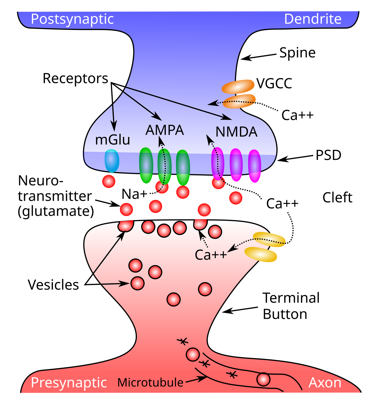
Figure 1: Schematic of a synapse, showing a presynaptic terminal button and the postsynaptic dendritic spine. Neurotransmitter (NT) is released into the synaptic cleft from the presynaptic side, and then binds to postsynaptic receptors in the postsynaptic density (PSD) of the spine, causing ion channels to open (e.g., sodium ions for excitatory AMPA channels), and thus activating the receiving neuron. NMDA receptors and voltage gated calcium channels (VGCC) allow calcium (Ca++) to enter the cell, triggering synaptic plasticity (learning). Metabotropic receptors such as mGluR do not open to allow ions to flow (as ionotropic ones do), and instead they trigger second-messenger cascades of reactions that can affect learning and other processes in the postsynaptic cell.
The synapse (Figure 1) has a lot of moving parts (introduced in neuron), any one of which could potentially be the critical factor in causing its overall efficacy to change. How many can you think of? The search for the critical factor(s) dominated the early phase of research on synaptic plasticity, and evidence for the involvement of a range of different factors has been found over the years, from the amount of presynaptic neurotransmitter released, to the number and efficacy of postsynaptic AMPA receptors, and even more subtle things such as the alignment of pre and postsynaptic components, and more dramatic changes such as the cloning of synaptic connections into multiple copies.
Our current understanding is that the dominant factor for synaptic plasticity in excitatory neurons in the neocortex is the number and efficacy of active postsynaptic AMPA receptors, which are those receptors that are embedded in the postsynaptic density (PSD) and exposed to the synaptic cleft so that glutamate can bind to them. The number of active AMPA channels is changed by a trafficking process that actively moves these channels between an internal “reserve” store and the PSD location. Pushing the AMPA receptor out into the PSD involves the processes of exocytosis while removing active AMPA receptors involves endocyctosis. These are routine processes that operate in every cell, on a remarkably rapid timescale: it is important to picture the cell membrane as a very pliable fatty substance that is easily penetrated.
To understand how LTP and LTD work at a biochemical level, we need to understand the chain of processes that ultimately regulate this AMPA trafficking process, which can be grouped into distinct stages:
• Induction is the first stage that starts everything off: the very first processes taking place on the timescale of milliseconds that are affected by the activity of the pre and postsynaptic neurons. This should determine whether the result will be LTP or LTD (or no change), and thus is the critical stage for determining the nature of the biological learning algorithm. As such, it is the focus of our discussion here.
• Expression is the next stage involving the initial changes in synaptic efficacy caused by the induction process. This involves both very rapid changes in the conductance of existing active AMPA receptors in the PSD, and the AMPA receptor trafficking changes that result in a change in the number of active AMPA receptors. In addition, rapid changes in the structure of the dendritic spine facilitate this trafficking process (Yang & Liu, 2022; Bosch et al., 2014; Matsuzaki et al., 2004). This structure is determined in part by actin polymer fibers, which are the same fibers involved in muscles!
• Early Maintenance (included in the term E-LTP) are the mechanisms that sustain these changes in expression over a period of seconds to minutes, and determine at this stage whether the changes will end up being short term plasticity (STP) by decaying away back to baseline, versus long term plasticity which persists through the next stage. These mechanisms must stabilize the changes in expression that took place, and it is critical to appreciate that the inside of a cell is a highly chaotic, active place with complex chemical reactions taking place continuously, so this early maintenance is not an automatic consequence of the initial expression changes. There is evidence that this early maintenance depends on the binding of CaMKII molecules to the NMDA receptors at a particular location on the NMDA receptor known as GluN2B (Bayer & Giese, 2025), and the consequent interaction with the structural changes in the shape of the spine (Yasuda et al., 2022).
• Late Maintenance (L-LTP) is the first stage that involves protein synthesis (the creation of new protein molecules), which requires the coordinated interaction of RNA and ribosomes, and generally takes around 60 minutes after induction in neurons. The new proteins are localized in the PSD (Tanaka et al., 2008) and also involve persistent changes in the actin fiber structures of the spine.
• Sleep processes: various changes happen during sleep to further stabilize and also update the synaptic changes that took place during the day. Throughout the body, major rebuilding and structural changes take place during sleep, and synapses in the brain are no exception. One popular idea is that the overall strength of synapses are rescaled in various ways during sleep (Abbott & Nelson, 2000; Tononi & Cirelli, 2003; Turrigiano, 2008; Torrado Pacheco et al., 2021), and there is also evidence for further sculpting of synaptic strengths (Li et al., 2017). All of these changes could contribute to well-documented changes in memory that occur after sleep (Klinzing et al., 2019). We found that incorporating a version of the mechanism described by Torrado Pacheco et al. (2021) results in significant improvements in the overall stability and efficacy of our models (see Slow weights).
The remainder of this page focuses on the mechanisms involved in the induction process, and how they interact with the remaining stages, without much further discussion of the details of these other stages (see above references for recent reviews). Remarkably, a central player in the induction process, CaMKII, provides a critical bridge to the subsequent stages (Bayer & Giese, 2025; Yasuda et al., 2022), albeit in a way that differs significantly from the original hypothesis proposed by John Lisman, which involved the ability of CaMKII to autophosphorylate itself and thus sustain a memory signal over time (Lisman, 1994). Instead, it now appears that memory maintenance is more like muscle strength maintenance, due to the critical involvement of actin fibers and structural changes in synaptic spines, which were originally proposed by Van Harreveld & Fifkova (1975).
NMDA and calcium influx
The NMDA receptor plays a central role in the induction of synaptic plasticity, because its opening allows calcium ions (\(Ca^{++}\)) to enter the postsynaptic spine, which then provides the initial trigger for a sequence of biochemical cascades that result in the changes to AMPA receptor conductance and number. Throughout the body, calcium ions play a similar role, and similar such biochemical cascades can be found in many different cell types. The voltage-gated calcium channels (VGCC) also conduct calcium, and play an additional important role, as do the metabotropic glutamate receptors (mGluR).
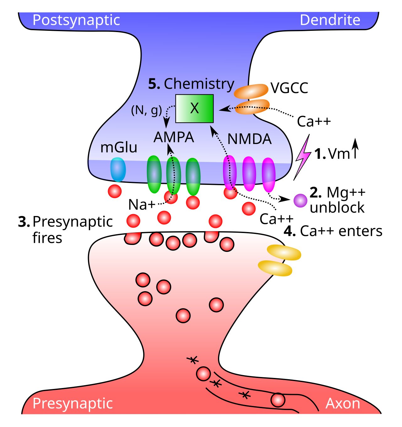
Figure 2: Critical steps in allowing calcium ions (Ca++) to enter postsynaptic cell via NMDA channels, inducing synaptic plasticity. 1. The postsynaptic membrane potential (Vm) must be elevated (from excitatory inputs to AMPA receptors across synapses of the postsynaptic neuron, and backpropagating action potential that comes back down the dendrite when the postsynaptic neuron fires). 2. Elevated Vm causes magnesium (Mg++) ions to be expelled from NMDA channel openings, thus unblocking them. 3. Presynaptic neuron fires an action potential, releasing glutamate. 4. Glutamate binds to NMDA receptors, causing them to open, allowing Ca++ to enter (only when also unblocked, per step 2). 5. The concentration of Ca++ in the postsynaptic spine drives a cascade of chemical processes (indicated by the X) that result in changes in AMPA receptor number and efficacy (N, g conductance), thereby changing the synaptic weight. Ca++ can also enter from voltage-gated calcium channels (VGCCs), which depend only on postsynaptic Vm levels, and not sending activity — these are weaker contributors to Ca++ levels.
Figure 2 shows the five critical steps involved in NMDA channel function:
1. The postsynaptic membrane potential (\(V_m\)) must be elevated, as a result of all the excitatory synaptic inputs coming into the cell. The most important contributor to this \(V_m\) level is actually the backpropagating action potential — when a neuron fires an action potential, it not only goes forward out the axon, but also backward down the dendrites (via active voltage-sensitive \(Na^+\) channels along the dendrites). Thus, the entire neuron gets to know when it fires — we’ll see that this is incredibly useful computationally.
2. The elevated \(V_m\) causes magnesium ions (\(Mg^{++}\)) to be repelled (positive charges repel each other) out of the openings of NMDA channels, unblocking them.
3. The presynaptic neuron fires an action potential, releasing glutamate neurotransmitter into the synaptic cleft.
4. Glutamate binds to the NMDA receptor, opening it to allow \(Ca^{++}\) ions to flow into the postsynaptic cell. This only occurs if the NMDA is also unblocked. This dependence of NMDA on both pre and postsynaptic activity was one of the early important clues to the nature of learning, as we see later.
5. The concentration of \(Ca^{++}\) in the postsynaptic spine drives the complex chemical reactions that end up changing the number and efficacy of AMPA receptors. Because these AMPA receptors provide the primary excitatory input drive on the neuron, changing them changes the net excitatory effect of a presynaptic action potential on the postsynaptic neuron. This is what is meant by changing the synaptic efficacy, or weight.
The fact that NMDA requires both pre and postsynaptic activity makes it essentially a direct mechanism for the Hebbian learning principle, which only increases synaptic connections when both the pre and postsynaptic neurons are active at the same time (Levy & Steward, 1979, Collingridge et al., 1983, Collingridge & Bliss, 1987). This is why most neuroscientists naturally assumed that Hebbian learning was the whole story (Cooper, 2005) — could it just be a coincidence that this complicated biological mechanism so directly implements a functionally motivated learning algorithm!?
\(Ca^{++}\) can also enter the postsynaptic cell via voltage gated calcium channels VGCC which are calcium channels that only open when the membrane potential is elevated. Unlike NMDA, however, they are not sensitive to presynaptic neural activity — they only depend on postsynaptic activity. This has important computational implications, as we discuss later. VGCCs contribute less to \(Ca^{++}\) levels than NMDA, so NMDA is still the dominant player.
Metabotropic glutamate receptors (mGluR) also play an important role in synaptic plasticity. These receptors do not allow ions to flow across the membrane (i.e., they are not ionotropic), and instead they directly trigger chemical reactions when neurotransmitter binds to them. These chemical reactions can then modulate the changes in AMPA receptors triggered by \(Ca^{++}\).
Calcium concentration determines LTP vs LTD
To determine the nature of the biological learning algorithm, we need to understand how this calcium signal coming from NMDA and VGCC channels results in LTP vs LTD depending on the nature of the pre and postsynaptic activity. For a long time, researchers focused mainly on LTP (which is generally easier to induce), but eventually they realized that both directions of synaptic plasticity are equally important for learning (Dunwiddie & Lynch, 1978; Lisman, 1989; Bear & Malenka, 1994).
A key development was the idea that the direction of plasticity depends on the amount of calcium that enters the cell, which is in turn a function of the overall synaptic activity as measured by the NMDA receptor. Because NMDA receptor opening depends on both the rate of presynaptic firing (for glutamate binding) and the rate of postsynaptic firing (for magnesium unblocking), the total amount of Ca++ entry should be a function of these variables times the overall duration of this activity:
\[ [Ca^{++}]_i = S R T \]
where \([X]_i\) is standard notation for the intracellular concentration of something.
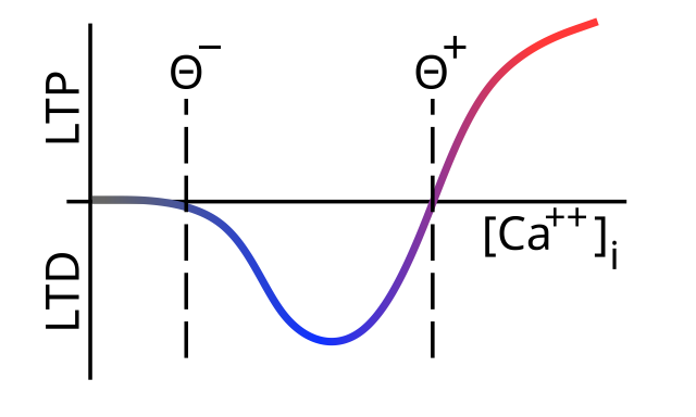
Figure 3: Standard account (after Lisman, 1989) for the direction of synaptic plasticity (LTP = increase, LTD = decrease) as a function of Ca++ concentration in the postsynaptic spine (accumulated over several hundred milliseconds). Low levels of Ca++ cause LTD, while higher levels drive LTP. Threshold levels indicated by theta values represent levels where the function changes sign.
Figure 3 shows the standard model, widely accepted to this day, for how the direction of change depends on the overall level of \([Ca^{++}]_i\) in the postsynaptic spine, accumulated over a few hundreds of milliseconds at least. Low levels drive LTD, while high levels produce LTP (Lisman, 1989; Bear & Malenka, 1994). Interestingly, the shape of the function in Figure 3 is essentially identical to one developed for a version of Hebbian learning known as the BCM rule in reference to the author’s initials (Bienenstock et al., 1982).
At a biochemical level, the basic model from Lisman (1989) was that the CaMKII kinase drives LTP, while a competing phosphatase pathway known as PP1 drives LTD, and that the differential sensitivities of these two pathways to the influx of \(Ca^{++}\) results in the shape of the curve shown in Figure 3. Specifically, PP1 is dominant at low calcium levels and thus drives LTD, while CaMKII dominates at higher levels and thus drives LTP as calcium levels increase. This original hypothesis is in need of significant updating in light of more recent changes in our understanding of CaMKII functioning, which we discuss in detail after the following discussion of STDP.
Spike timing dependent plasticity
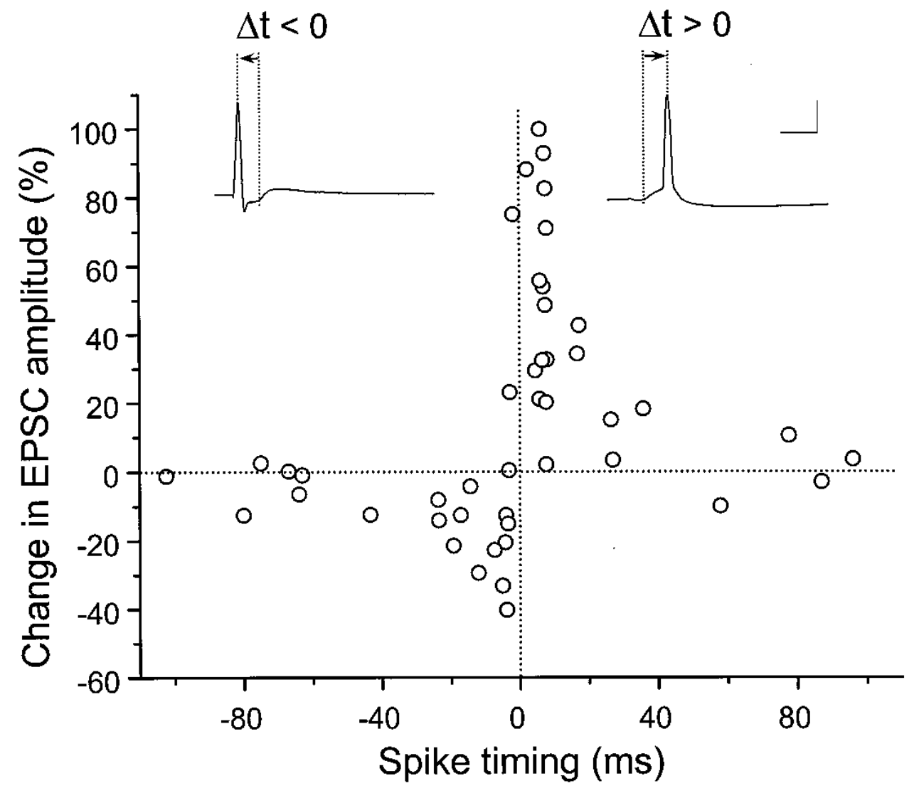
Figure 4: Spike timing dependent plasticity demonstrated in terms of temporal offset of firing of pre and postsynaptic neurons. If post fires after pre (Delta t > 0, right side), then the weights go up, and otherwise (Delta t ≤ 0, left side) they go down. This fits with a causal flow of information from pre to post. Reproduced from Bi and Poo (1998).
It is fair to say that the finding of spike-timing dependent plasticity (STDP) by Bi & Poo (1998) has been the single most impactful experiment in the field since the original Bliss & Lomo (1973) finding, with more than 30,000 papers containing that term on google scholar (in quotes!) and over 5,000 citations of the original paper. Figure 4 shows the key result: synaptic learning seems to obey a simple, appealing causality principle, such that LTP occurs when the presynaptic neuron fires before the postsynaptic one, and vice-versa for the opposite order. This intuitively appealing logic has proven especially attractive to a large number of computational modelers, who have extensively employed STDP-like mechanisms (e.g., Kheradpisheh et al., 2018; Diehl & Cook, 2015).
A simple version of the causal argument suggests that if a given pre neuron actually played a role in driving the post neuron to fire, then it must necessarily have fired in advance of it, and according to the STDP results, its weights will increase in strength. Meanwhile, pre neurons that have no causal role in firing the postsynaptic cell will have their weights decreased.
The vast literature on STDP has since revealed a tremendous amount of variability in whether you get LTP, LTD, or neither, depending on many different variables (Debanne & Inglebert, 2023), including a dependency on the non-naturalistic calcium and magnesium concentrations used in the in vitro experiments (see in activo). Furthermore, the original experiment required highly specific and artificial stimulation conditions involving single isolated pairs of spikes separated by a full second of no activity, with many repetitions of a given such timing pattern required to produce the STDP results.
Critically, when even slightly more complex spike trains were used, the results were not consistent with a simple application of the classical STDP plasticity function, and instead reveal considerable more complexity in the nature of the underlying mechanism (Froemke & Dan, 2002; Froemke et al., 2006; Urakubo et al., 2008). A similar conclusion was reached by Shouval et al. (2010), who argue that a version of the BCM (Bienenstock et al., 1982) mechanism provides a better overall account. We replicate this conclusion using the biologically detailed model of Urakubo et al. (2008), which produces the classic STDP curve under the original artificial stimulation conditions, but more generally produces BCM-like functions, as described below and in the urakubo08 simulation. In short, this work suggests that STDP is a special case consequence of the underlying synaptic plasticity mechanisms, rather than a good description of what they accomplish in general.
Finally, a consideration of the natural patterns of neural activity in the awake, behaving neocortex raises fundamental questions about the causal logic behind the STDP theory in the first place. Specifically, there is considerable persistent ongoing neural activity at all levels of the neocortex, which has been shown to strongly contextualize the responses of neurons to incoming stimuli, for example (Arieli et al., 1996). Thus, contrary to the strictly feedforward causal direction envisioned by models based on STDP, activity in the brain does not have a simple reliable causal arrow due to the extensive bidirectional connectivity.
Biochemical cascades of kinases and phosphatases
To better understand how LTP and LTD work in the brain, we need to understand more about the complex biochemical cascades that are triggered by the influx of calcium into the postsynaptic spine. We first discuss what can now be termed the classical model based on standard principles of biochemistry and a number of experimental results and theories that were developed through the 2010s. Then, we discuss the newer structural model that provides a better overall fit to the available data (Bayer & Giese, 2025).
There are two major players in these biochemical cascades (in addition to the calcium itself):
• Kinases are protein structures that act as a catalyst for promoting other chemical reactions. They are typically activated by the binding of other substances at specific binding sites, which are identified by an amino acid and a number indicating the distance in amino acids from the N terminal end of the protein chain (e.g., Thr286). Kinases typically act to phosphorylate other molecules, which then activates them so they can bind to other molecules, or perform some other function. Phosphorylation means adding a phosphate group (compounds involving \(PO_4\)), and you may remember from high school biology that ATP is the energy carrier in cells, where the P in ATP is phosphate (adenosine triphosphate). Phosphate = energy or activation in a very general sense.
• Phosphatases play an opposing role to kinases: they act to dephosphorylate other molecules, and thus typically deactivate or inhibit them. However, there are also cases where dephosphorylation of one site will actually enable binding at other sites, due to corresponding changes in the overall shape of the molecule for example. Thus, there is no hard-and-fast rule that says a kinase is always excitatory or activating and a phosphatase is always inhibitory or de-activating.
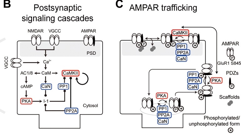
Figure 5: Diagram of the chemical processes included in the Urakubo et al. (2008) model, which are driven by Ca++ influx from NMDA and VGCC receptors. Ca++ binds to Calmodulin (CaM) which then directly and indirectly binds to two key kinases: CaMKII and PKA (shown in red). Opposing these kinases are three phospatases (shown in blue), CaN (calcineurin), PP1 (protein phospatase 1) and PP2A. Per the Lisman (1989) model, CaMKII drives LTP by promoting the insertion of AMPA receptors into the postsynaptic density (PSD) where they are active, and PP1 drives LTD by promoting the endocyctosis of AMPA receptors into an inactive reserve pool. Reproduced from Urakubo et al, 2008.
Figure 5 shows a “standard model” of the cascade of chemical processes that are triggered by \(Ca^{++}\) influx via NMDA and VGCC receptors, reflecting the general level of understanding through the first two decades of the 2000s. A detailed biochemical model based directly on the empirically determined rate constants for all of these chemical processes was developed by Urakubo et al. (2008), a replication of which is available in urakubo08 simulation to explore. This model includes the AMPA receptor trafficking mechanisms, so we measure changes in synaptic efficacy in this model directly in terms of the number of AMPA receptors in the PSD after these slower processes have had a time to take effect. Although this model is missing some critical newer elements, it nevertheless provides a useful tool for understanding how these chemical processes operate in detail, with many well-validated components that can be used in updated models. Furthermore, it does account for a wide range of existing data as we’ll see.
The cascade starts with CaM (calmodulin) which is the primary direct binding target for \(Ca^{++}\), which activates this protein by inducing a change in its shape that enables it to bind to other targets. Activated CaM then activates CaN (calcineurin, a phosphatase), and the CaMKII kinase (calcium/calmodulin-dependent protein kinase 2), which we’ve mentioned several times already, and is clearly the single most important protein in the complex network of interacting players involved in synaptic plasticity. Further downstream in the cascade is PP1 (protein phosphatase 1), which is a major opponent to CaMKII according to the standard LTP / LTD model of Lisman (1989). Another kinase named PKA (protein kinase A) plays a role in inhibiting PP1 activation, and it is also several steps downstream of CaM activation.
The fact that CaMKII is directly activated by CaM, whereas PP1 is a few more steps downstream of CaM, creates a significant difference in their temporal dynamics, where CaMKII is activated more quickly and PP1 takes longer to activate as we’ll see. These dynamics are based on prior models that used detailed measurements of the underlying chemical processes (Bhalla & Iyengar, 1999; Kuroda et al., 2001; Doi et al., 2005).
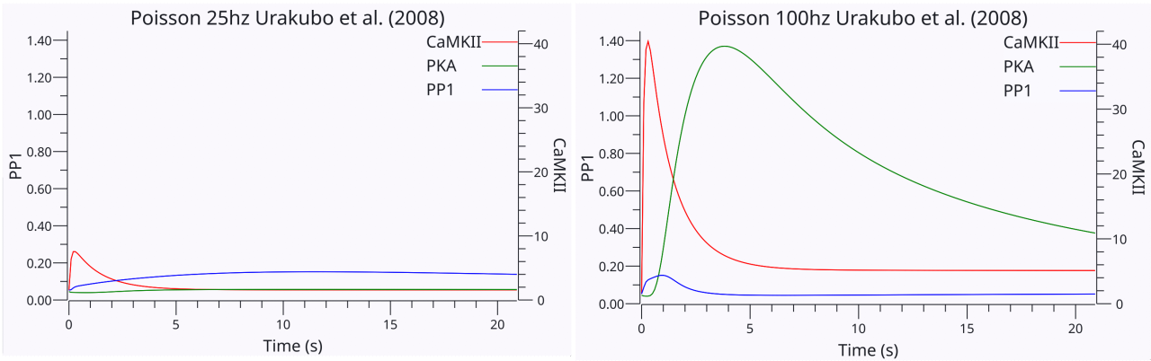
Figure 6: Dynamics of CaMKII, PKA, and PP1 in the Urakubo et al. (2008) model, in response to 200 ms of Poisson random spike trains of 25hz (left) or 100hz (right) pre and post activity, with a 20s post-activity period where the slower chemical proceses and AMPA trafficking settle out. The 25hz activity results in LTD (endocytosis of AMPA receptors), driven by the higher sustained activity of PP1 relative to CaMKII. PKA does not get sufficiently active to inhibit the PP1 pathway. Also note the much longer activity of PP1 vs. CaMKII, which builds on the longer activation of CaN (not shown). In the 100hz case, the increased initial activity is sufficient to persistently activate CaMKII and results in LTP instead of LTD. Furthermore, PP1 ends up being inhibited via the strongly activated PKA pathway.
To see how these chemical processes operate in response to pre and post spiking activity, Figure 6 shows the timecourses of CaMKII, PKA, and PP1 in response to 200 ms of Poisson random spiking our replication of the Urakubo et al. (2008) model. You can see that PP1 has a longer overall timecourse for the 25Hz random activity case in the first figure, and it ends up out-competing the transient activity of CaMKII, thus resulting in LTD (endocytocis of AMPA receptors from the postsynaptic density, PSD). In the 100hz random activity case in the second figure, CaMKII is sufficiently activated initially to remain active over time, and the stronger activation of the PKA pathway via the higher levels of calcium and CaM activation ends up inhibiting the PP1 pathway. This results in LTP (expression of the AMPA receptors in the postsynaptic density).
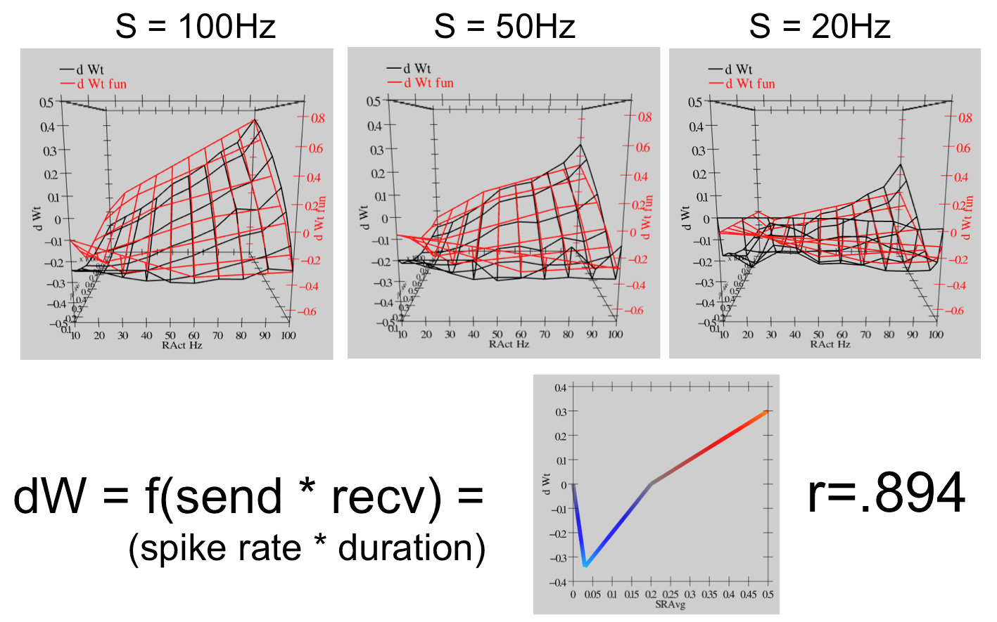
Figure 7: The overall behavior of the Urakubo et al. (2008) model in response to spike trains with different frequencies and durations is shown in the black mesh 3D plot, where the depth Z axis is duration of spiking in portions of a second (0-1), the X axis is receiver firing frequency in Hz, and the different plots show different sender firing frequencies. All of this data can be captured at a high level of accuracy (Pearson’s r=0.894) with a simple piecewise-linear function of the total “synaptic activity” which is the product of sending and receiving firing rate times the duration. This function, shown at the bottom right and as applied to the data in the red mesh plots, has a qualitative BCM-like shape, although in a more linearized form, with moderate levels of Ca++ producing LTD and higher levels producing LTP.
We further tested the extent to which this model replicates the BCM-like curve shown in Figure 3, which is predicted by by the Lisman, 1989 CaMKII vs PP1 model, by systematically probing with spike trains of different frequencies and durations. The results, shown in Figure 7, confirm that this model does exhibit BCM-like behavior across a wide range of activity parameters, with a simple linearized version of the BCM function capturing a high level of variance (r=0.894). You can replicate this data yourself in the urakubo08 simulation.
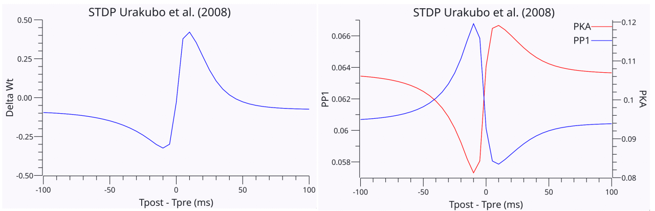
Figure 8: The Urakubo et al. (2008) model also replicates the STDP results of Bi and Poo (1998) using the standard protocol of single pairwise spikes at 1s intervals. Obtaining this behavior from the model required modifying the effective decay (inactivation) constant of the NMDA channels, so that glutamate binding from presynaptic release (i.e., pre before post, in the causal direction) resulted in a significant reduction in the rate of inactivation relative to glutamate unbound NMDA channels. Thus, the pre-before post direction (right side of curve) had much higher levels of Ca++ influx due to the longer open time of the NMDA channels. The resulting level of Ca++ influx and CaM activation mirrored the Delta-Wt learning curve shown in the first panel. Consistent with earlier figures, PP1 drives LTD for post-before-pre timings (left side of the curve), but CaMKII and PKA dominate on the pre-then-post side.
The Urakubo et al. (2008) model can also replicate the Bi & Poo (1998) STDP results as shown in Figure 8, but only with a very specific modification that directly supports the STDP phenomenon. Specifically, they implemented allosteric dynamics where the binding of glutamate to the NMDA receptor alters its rate of inactivation by a factor of 100, going from 2.3 ms (unbound) to 230 ms (bound). The term allosteric refers to any modulation of protein function by binding to a different site than the one directly involved in a reaction (which is very common), so here glutamate is modulating the inactivation reaction which is driven by CaM binding, as a result of calcium entry. Thus, if the presynaptic neuron fires first, then the glutamate binding to the NMDA receptor causes it to stay open significantly longer and allow a high LTP-level of calcium to enter. Conversely, if postsynaptic fires before presynaptic, the NMDA receptor has the much shorter inactivation time constant and only an LTD-level of calcium enters.
Although this hypothesized mechanism is supported by additional data, it is unclear if it can be considered a well established property of the NMDA channel (e.g., it is not mentioned specifically in a recent thorough review of the inactivation mechanisms of NMDA channels; Iacobucci & Popescu, 2024). This raises the question as to whether other mechanisms missing from the Urakubo et al. (2008) model may instead provide the relevant differentiator behind the STDP phenomenon. Nevertheless, even in the extensive additional results from the Urakubo et al. (2008) model on more complex versions of STDP-like paradigms, there is ample evidence that the simple causal STDP story does not provide an adequate explanation of the nature of synaptic plasticity, as we discussed previously.
Structural role of CaMKII binding to NMDA GluN2B
The “classical” model of CaMKII functioning described above has been challenged by a number of findings that significantly contradict its central premises. In its place, a new understanding of how CaMKII functions is emerging, based on a structural binding mechanism, rather than the standard catalytic model of what kinases typically do (Bayer & Giese, 2025). Specifically, binding site Thr286 on CaMKII binds to the GluN2B location of the NMDA receptor, and this binding appears to be critical for driving the subsequent synaptic plasticity stages that CaMKII is involved in (Tullis & Bayer, 2023; Chen et al., 2024).
This is a structural account because it is the physical location of the CaMKII attached to the NMDA receptors, not its catalytic activation, that is the essential causal step. Nevertheless, many of the other processes from the classical model remain relevant. For example, the very first step of Ca++-activated CaM binding to activate CaMKII, which occurs at Thr286, is required for it to bind to GluN2B in the first place, so only CaMKII that has been activated by calcium entry will engage in this structural binding.
By contrast, this same Thr286 binding site on the CaMKII protein was hypothesized to be critical for its ability to sustain a memory trace through autocatalytic phosphorylation (Lisman, 1994). Specifically CaMKII can form complexes of 12 protein elements all bound together via this binding site, and once this occurs, the theory was that it would keep the CaMKII in a stable state of kinase activation over an indefinite period, thereby providing the long-term memory maintenance signal. However, accumulating data strongly suggests that CaMKII is only active transiently when activated Ca++-CaM molecules are around, so it therefore does not seem plausible as a long-term memory signal (Bayer & Giese, 2025).
The new structural model thus provides a way for this transiently activated CaMKII to nevertheless drive a longer-lasting memory cascade: transient activity is needed for the structural GluN2B binding. Furthermore, the result of this structural binding is likely similar to what Lisman (1994) originally proposed, which is a stabilization of multiple CaMKII complexes bound together at the Thr286 site. What is new is that this CaMKII is now concentrated around the NMDA receptors in the PSD, which puts them physically in an opportune place to trigger multiple further events that result in AMPA exocytosis, actin fiber reorganization, and eventual protein synthesis at the PSD.
Structural LTD mechanisms: DAPK1 and Thr305/306
One critical feature of the GluN2B structural binding mechanism of CaMKII function is that it provides an entirely different way of understanding how the switch between LTD and LTP might operate: anything that interferes with CaMKII binding to GluN2B should result in LTD, while anything that facilitates it should result in LTP.
One important mechanism that interferes with CaMKII GluN2B binding and results in LTD is the competitive binding of DAPK1 (death-associated protein kinase 1) to this same binding site (Goodell et al., 2017; Goodell et al., 2021; Cook et al., 2021; Tullis & Bayer, 2023). DAPK1 is structurally and functionally similar to CaMKII (Bayer & Schulman, 2019), but is more widely expressed and as the name suggests, it is typically involved in triggering cell death (apoptosis). But in neurons, it appears to have a more specific function.
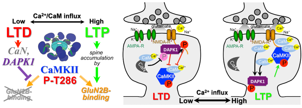
Figure 9: Competition between DAPK1 and CaMKII for binding to the GluN2B binding location on NMDA receptors determines the direction of synaptic plasticity between LTP and LTD, based on the results from Goodell et al. (2017). Consistent with the initial stages of the standard model discussed above, the phosphatase Calcineurin (CaN) is preferentially activated by lower levels of Ca++ entry. In this new picture, it then dephosphorylates one site on DAPK1 (S308), which then disinhibits CaM binding at another site (S1303), which in turn promotes binding of DAPK1 to the GluN2B location on NMDA receptors at Thr286. This competitive GluN2B binding prevents CaMKII from binding, and leads to LTD. However, higher levels of Ca++ influx enable CaMKII to win this competition and leads to LTP via GluN2B binding.
Specifically, as shown in Figure 9, Goodell et al. (2017) found that DAPK1 binds to GluN2B when a different binding site (S308) on DAPK1 has been dephosphorylated by activated CaN (calcineurin) (i.e., this is an allosteric mechanism where dephosphorylation results in activation). As shown in Figure 5, CaN is directly activated by Ca++-activated CaM, so DAPK1 is one step removed from direct Ca++ influx, while CaMKII is directly activated by CaM. Using the standard BCM-like logic, Goodell et al. (2017) argued that low levels of calcium favor CaN and DAPK1 activation for preferential binding to the GluN2B site, while higher levels favor CaMKII in the competition over this one binding site. When CaMKII wins, LTP results, but when DAPK1 wins, LTD results.
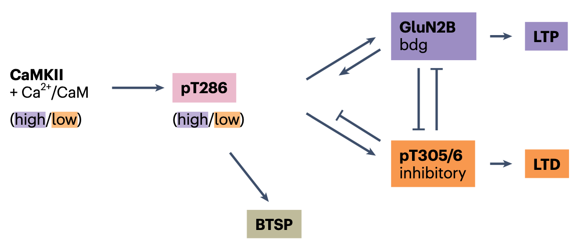
Figure 10: Interactions between the GluN2B binding and phosphorylation of site Thr305/306 of CaMKII in driving the induction of either LTP or LTD, according to the structural model of Bayer & Giese (2025). When Thr305/306 is activated, binding to GluN2B is inhibited. This operates in addition to, and in cooperation with, the DAPK1 competition for the GluN2B binding site shown in the previous figure. Reproduced from Bayer & Giese (2025).
In addition to competition from DAPK1 binding, the activation of a different binding site on the CaMKII protein, Thr305/306 (nearby to the Thr286 site) also inhibits the binding of CaMKII to the GluN2B site (Cook et al., 2021; Figure 10). The results indicate that both this Thr305/306 and DAPK1 activation are required to induce LTD, as blocking either of them prevents LTD. The activation of the Thr305/306 site occurs via binding by other CaMKII proteins (i.e., autocatalyzation or autophosphorylation), and to make things even more complicated, this autocatalyzation requires initial activation of Thr286 by Ca++-CaM, which had previously been shown to be essential for both LTP and LTD (Coultrap et al., 2014).
Of particular relevance to the temporal derivative mechanism that underlies the kinase algorithm, the temporal dynamics of this Thr286 and Thr305/306 autophosphorylation result in a strong differential sensitivity to fast vs slower patterns of activation by Ca++-CaM binding. Specifically, as explained in Cook et al. (2021), there is a fast initial response to Ca++-CaM activation at Thr286 that promotes the binding of multiple CaMKII molecules together at that site, which if sustained would result in subsequent binding to the GluN2B site on NMDA, and result in LTP. However, the CaM molecules will dissociate from the Thr286 site relatively quickly, and when this occurs, the CaMKII molecules instead tend to bind together at the nearby Thr305/306 site, which inhibits GluN2B binding.
Thus, the relative balance of Thr286 vs. Thr305/306 activation appears to be ideally situated to detect differences in the overall rate of synaptic activity. In particular, a pattern of synaptic activity that starts out strong but then drops down to a lower rate would seem to be ideal for activating the Thr305/306 binding, resulting in LTD. This is consistent with the results from Jang et al (2026) showing LTD for this pattern of activity, which corresponds computationally to a prediction error: an initially strong expectation (high synaptic activity) was not confirmed in the outcome, resulting in a subsequent drop in synaptic activity.
In summary, both the DAPK1 and this Thr305/306 overall dynamic of different pathways with different effective time constants competing with each other to determine whether LTP or LTD occurs is the hypothesized mechanism for the temporal derivative mechanism that the kinase algorithm uses to implement error-driven learning. A major goal of future research is to adapt the urakubo08 simulation to include the above structural GluN2B mechanisms, to determine more precisely their temporal response profile.
More generally, a critical lesson from all of the research on synaptic plasticity is that the standard paradigms used for induction are relatively limited, and specifically do not include the kind of rapid temporal differences required for the temporal derivative mechanism. Thus, as the results from Jang et al (2026) demonstrate, this research may be overlooking the most important conditions for driving LTP vs. LTD, by focusing either on simple bursts of steady-state activity at very different frequencies, or on the highly artificial STDP paradigm.
Temporal eligibility traces
In addition to rapid temporal dynamics on the timescale of a few 100 milliseconds, there are longer timescales that are likely to be important for shaping synaptic plasticity. For example, consider the time interval between when an action is performed, and when the subsequent reward or punishment associated with that action is experienced. Depending on the situation, this time interval could span anywhere from a second up to hours, days or even longer.
The initial synaptic activity associated with the performance of the action is thought to initiate an eligibility trace for the relevant synapses that allows them to be modified by the activity that occurs during the subsequent outcome, thereby establishing a critical cause-and-effect relationship across these two points in time.
Recent data from neurons in area CA1 of the hippocampus shows evidence of learning contingencies that extend across the shorter end of these behavioral timescales, which has been termed behavioral timescale synaptic plasticity (BTSP; Bittner et al., 2015; Bittner et al., 2017; Milstein et al., 2021; Fan et al., 2023; Magee, 2026). Specifically, these neurons can exhibit rapid changes in their spatial tuning as a result of elevated dendritic plateau potentials, which can be separated from the relevant presynaptic activity by several seconds.
These plateau potentials in BTSP occur as a result of synaptic inputs from the entorhinal cortex, and are associated with goal outcomes. Thus, they are thought to function like a “third factor” training signal that indicates the consequences of actions (Gerstner et al., 2018). Other such third factors include neuromodulators such as dopamine, acetylcholine, norepinepherine, and serotonin, all of which have been shown in various ways to modulate learning, and are associated with reward and punishment outcomes in reinforcement learning paradigms. Two of these neuromodulators, norepinepherine and serotonin, where found to have a delayed BTSP-like effect on plasticity in the cortex, for example (He et al., 2015; Shouval & Kirkwood, 2025).
Although an understanding of the underlying mechanisms behind these phenomena is only recently becoming available, initial indications are that it involves all of the same mechanisms discussed above, including a central role for CaMKII and likely the structural Thr286 binding (Xiao et al., 2023; Fan et al., 2023; Jain et al., 2024; Bayer & Giese, 2025). The persistent presence of the structurally bound CaMKII likely enables it to span the relevant temporal intervals to connect the presynaptic input events with the later outcome-based signals.
One interesting property of the CA1-based BTSP mechanism is that there is a negative feedback loop that inhibits further learning, based on a specific class of inhibitory interneurons that receives excitation from CA1 pyramidal neurons and projects directly into the distal dendrites where the plateau potentials arise (Turi et al., 2019; Rolotti et al., 2022; Campbell et al., 2026). This inhibitory feedback loop mechanism prevents runaway synaptic plasticity. Furthermore, the BTSP form of plasticity appears to decay relatively rapidly (Vaidya et al., 2025), consistent with the idea that it represents a fast-but-transient form of plasticity for adapting output responses to the current behavioral contingencies (Magee, 2026; see also the stability-plasticity dilemma).
A similar BTSP-like mechanism has recently been described in layer 5 pyramidal neurons in neocortex (Yaeger et al., 2025; Xiao et al., 2025). These neurons provide the primary cortical output signals, and are thus appropriate targets for a rapid but transient output mapping process, similar to that used in reservoir computing. Like the CA1 pyramidal neurons, these layer 5 neurons also have a prominent distal dendritic tuft, in layer 1, where the plateau potentials were found. Also, some of these studies specifically looked for and did not find evidence of these plateau potentials in other neocortical pyramidal neurons (Yaeger et al., 2025), suggesting that these are unique to these deep output layer neurons.
The driving target signal for plasticity in these layer 5 neurons remains unclear (Magee, 2026), but we do know that these neurons receive extensive thalamic input targeting the distal dendritic tuft in layer 1. The thalamic projections that target layer 1 are generally of the matrix type, which means they typically have broad axonal arbors targeting many different neurons across multiple cortical areas. Thus, they appear ideally situated to provide the same kind of very broad signal as the neuromodulators like dopamine.
One class of such projections arises specifically from motor-associated thalamic areas (see frontal thalamus), which could then provide a way for widespread cortical output learning to optimize the alignment of cortical representations for the current behavioral outputs. Specifically the ventral anterior (VA) nucleus projects to essentially the entire neocortex, and receives inputs from layer 5 neurons in motor cortical areas, and is also under disinhibitory control from the basal ganglia (Phillips et al., 2021; Xiao et al., 2009; Kuramoto et al., 2015; Economo et al., 2018).
Another earlier literature on these temporal bridging mechanisms focused on synaptic tags that could persist for an hour or more, based on the distinct mechanisms involved in late maintenance of plastic changes as mentioned above (Morris & Frey, 1997; Reymann & Frey, 2007; Redondo & Morris, 2011). Given the need to connect the induction events with these later protein-synthesis dependent mechanisms, it would seem that this kind of temporal bridging is already a necessary feature of the post-induction chemical cascades.
The kinase algorithm used in Axon includes a parameter that allows synaptic activity traces to persist over time, and the Rubicon framework depends critically on such trace mechanisms in the basal ganglia, amygdala and other areas, to learn connections across behaviorally relevant timescales. At a computational level, Bellec et al. (2020) showed how a form of error backpropagation through time could be accomplished using eligibility traces of this same form.