
Cerebellum
The cerebellum is a system of highly specialized neural elements many of which are unlike those found in any other part of the brain. The precise function of all this specialized machinery, which is replicated across many parallel channels and connects with most of the sensory and motor pathways in the brain, has been tantalizingly elusive since the seminal publication of many of its unique features (Eccles et al., 1967).
Although it is widely considered to be a motor control system, the cerebellum is anatomically and evolutionarily a sensory system, albeit one that is tightly interconnected with the motor system. It is clearly involved in learning, but exactly what and how it learns remains a topic of active debate.
We adopt a specific implementation of the adaptive filter model (Fujita, 1982; Dean et al., 2010; Cullen, 2023), which filters out the predicted sensory signals generated by one’s own motor actions (i.e., sensory reafferance) to provide a more stable and useful basis for motor behavior. These sensory predictions are also be used as a forward model (Miall & Wolpert, 1996; Bastian, 2006; Shadmehr et al., 2010; Tanaka et al., 2020), to anticipate the sensory consequences of motor actions, in driving further actions that are therefore more coordinated. These are consistent with, but significantly more specific than, the more general error-driven learning models originally proposed by Marr (1969), Albus (1971), and Ito (1972), which remain the most widely discussed (Raymond & Medina, 2018; Shadmehr, 2020).
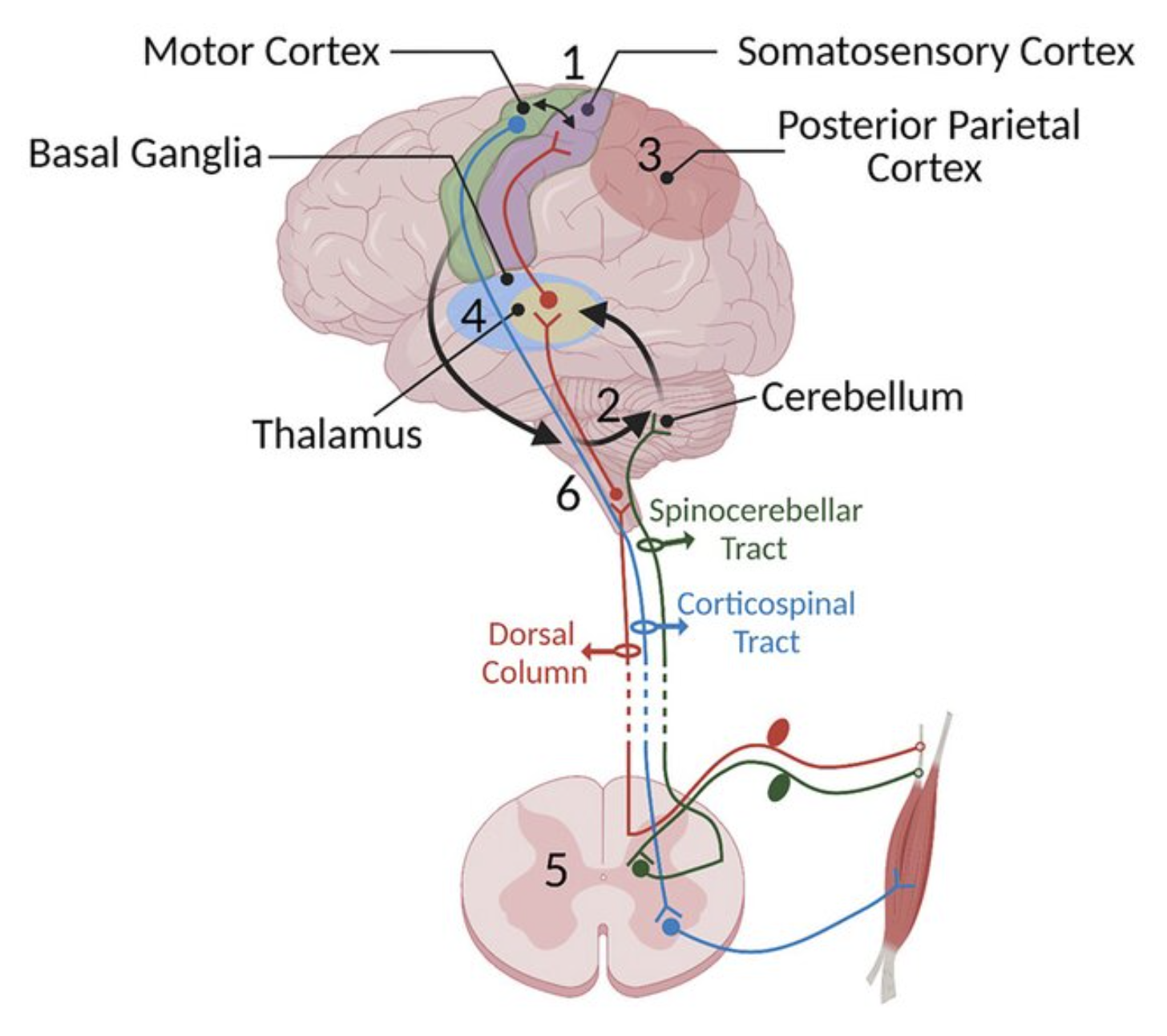
Figure 1: The location of the cerebellum in relation to other elements of the motor system. 5 is the spinal cord, and 6 is the brainstem. From Asan et al (2022).
The cerebellum is located at the base of the brain, forming a kind of outgrowth of the midbrain (Figure 1). It receives inputs from all of the major motor-associated areas of the brain, including the sensory-motor areas of the spinal cord and midbrain, the basal ganglia, and, via the pons or pontine nuclei, motor-associated cortical areas including primary motor and somatosensory cortical areas (M1 and S1). It also receives a wide range of other sensory signals, including a major projection from the vestibular system.
The cerebellar outputs, from the deep cerebellar nuclei (DCN or just CN in more recent terminology), project widely to many areas of the brain, including a major driving projection to the VL thalamus from the more lateral CN areas, which can provide a training signal to primary motor cortex. The possibility that the cerebellum is critical for early learning, but can then transfer this learning by training other structures including the cortex, is supported by the differential severity of early developmental cerebellar lesions (Beuriat et al., 2020).
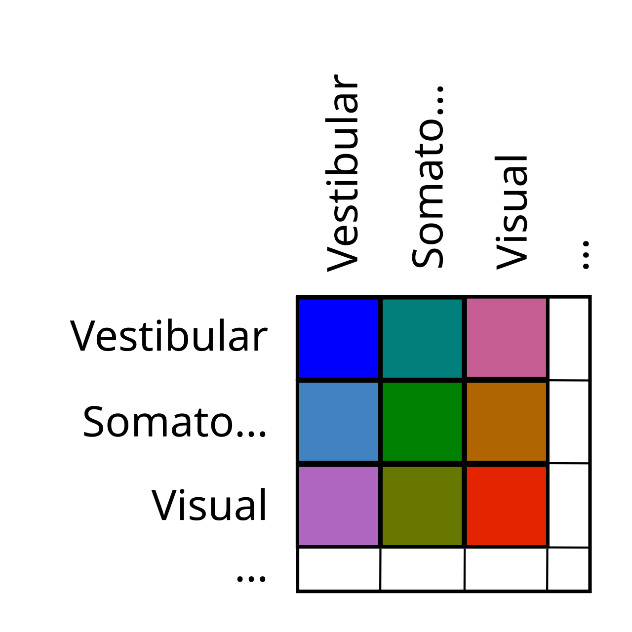
Figure 2: The cerebellum is organized like an outer product, representing many different combinations of sensory signals, including motor efferents.
Unlike the basal ganglia, neocortex, and thalamus, the cerebellum does not have a simple topographic organization that can be easily labeled in a figure. Instead, the primary function of the cerebellum is to integrate and compare signals across different modalities, so it is organized somewhat like an outer product across its various inputs, with each region comparing one sensory input against another in all manner of combinations (Figure 2).
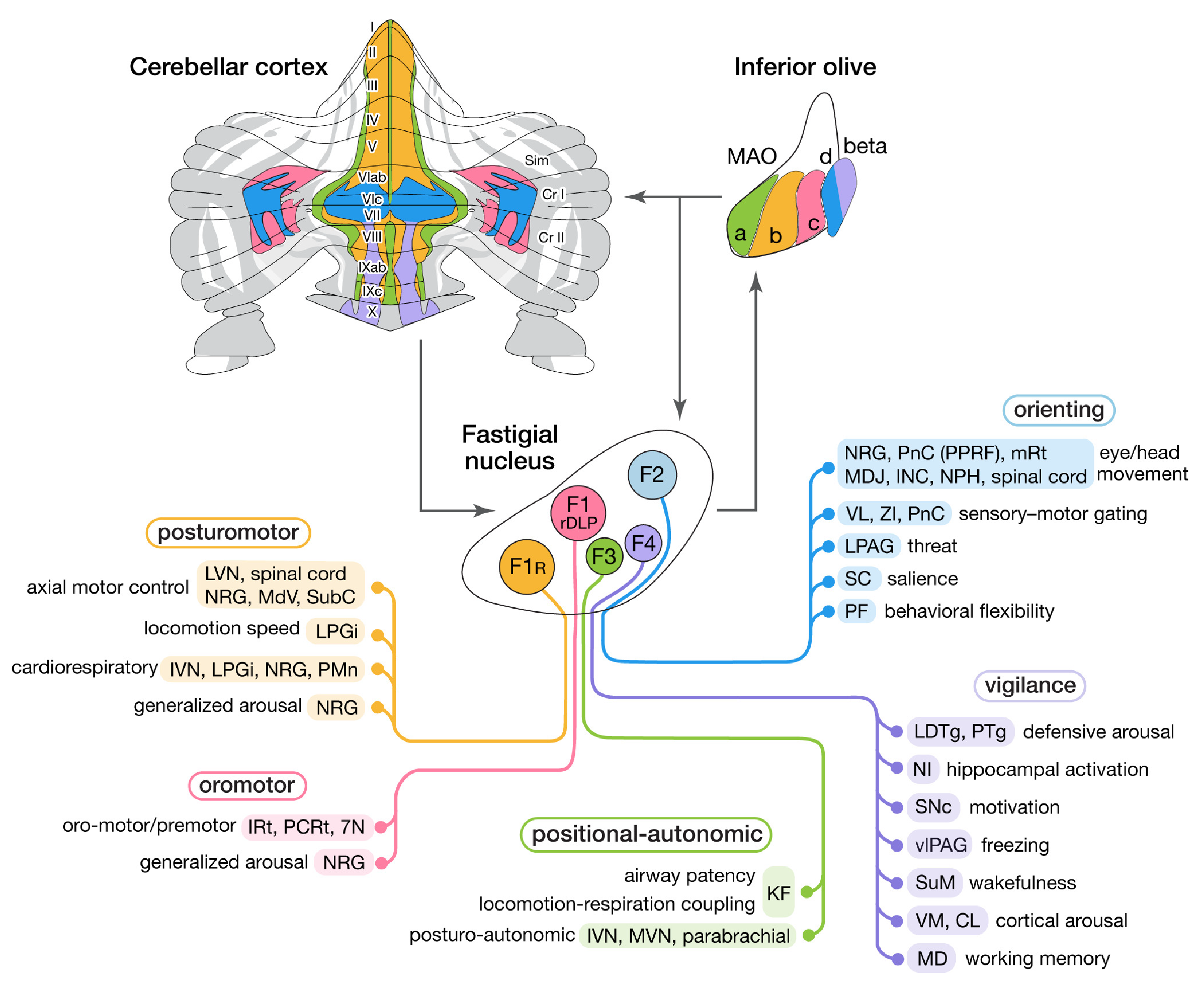
Figure 3: The different output pathways of the fastigial cerebellar nucleus, which integrate a range of different sensory signals to provide useful signals for essentially all major subcortical brain systems, consistent with this nucleus being the most evolutionarily ancient in the cerebellum. Much of the cerebellar cortex that it interconnects with is in the vermis, which is the midline portion of the cerebellum. 7N: facial nucleus; CL: centrolateral thalamic nucleus; INC: interstitial nucleus of Cajal; IRt: intermediate reticular nucleus; IVN: inferior vestibular nucleus; KF: Kölliker-Fuse nucleus; LDTg: laterodorsal tegmental nucleus; LPAG: lateral periaqueductal gray; LPGi: lateral paragigantocellular nucleus; LVN: lateral vestibular nucleus; MD: mediodorsal thalamic nucleus; MDJ: mesodiencephalic junction; MdV: medullary reticular nucleus, ventral; mRt: mesencephalic reticular formation; MVN: medial vestibular nucleus; NPH: nucleus prepositus hypoglossi; NI: nucleus incertus; NRG: nucleus reticularis gigantocellularis; PCRt: parvocellular reticular nucleus; PF: parafascicular thalamic nucleus; PMn: paramedian reticular nucleus; PnC: pontine reticular nucleus, caudal; PPRF: paramedian pontine reticular formation; PTg: pedunculotegmental nucleus; Sim: simplex lobule; SC: superior colliculus; SNc: substantia nigra, pars compacta; SubC: subcoeruleus nucleus; SuM: supramammillary region; VL: ventrolateral thalamic nucleus; vlPAG: ventrolateral periaqueductal gray; VM: ventromedial thalamic nucleus; ZI: zona incerta. From Fujita et al (2020).
Figure 3 shows an example of this outer-product like organization (Fujita et al., 2020), associated with the fastigial nucleus (FN), which is the evolutionarily most ancient of the cerebellar nuclei (Zhang et al., 2016). The FN is interconnected mostly with the medial vermis region of the cerebellar cortex, which integrates different signals including vestibular, somatosensory and interoceptive signals of relevance to different core survival-relevant systems.
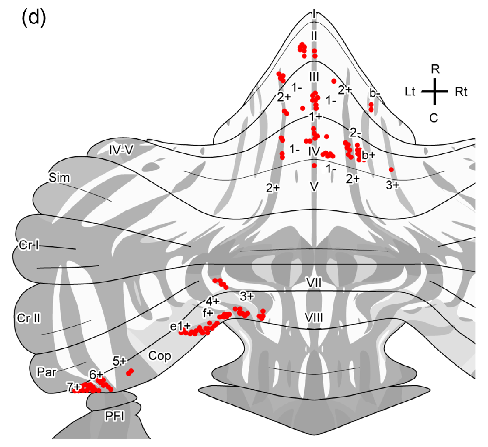
Figure 4: The projection terminals of a single neuron in the pons, conveying a specific sensory input signal into the cerebellar cortex. This one signal is spread widely across many areas, consistent with the outer-product organization of the cerebellum (e.g., this is one row or column in the outer-product matrix). The dark vs. light grey tones indicate the zebrin-based regions (dark) that have distinct gene expression profiles and other factors. From Biswas et al (2019).
Another representation of the thorough mixing of sensory signals in the cerebellum is shown in Figure 4, plotting the terminal zones of the axon of a single mossy fiber from a neuron in the pons (Biswas et al., 2019). This one sensory signal is distributed across many areas of the cerebellar cortex, and many such neurons also project down to the cerebellar nuclei as well. There are around 43 million total neurons in the cerebellum of the adult mouse (Surchev et al., 2007), reflecting the huge combinatorial explosion of the outer-product organization, along with a major additional multiplier by the numerous granule cells in the cerebellar cortex.
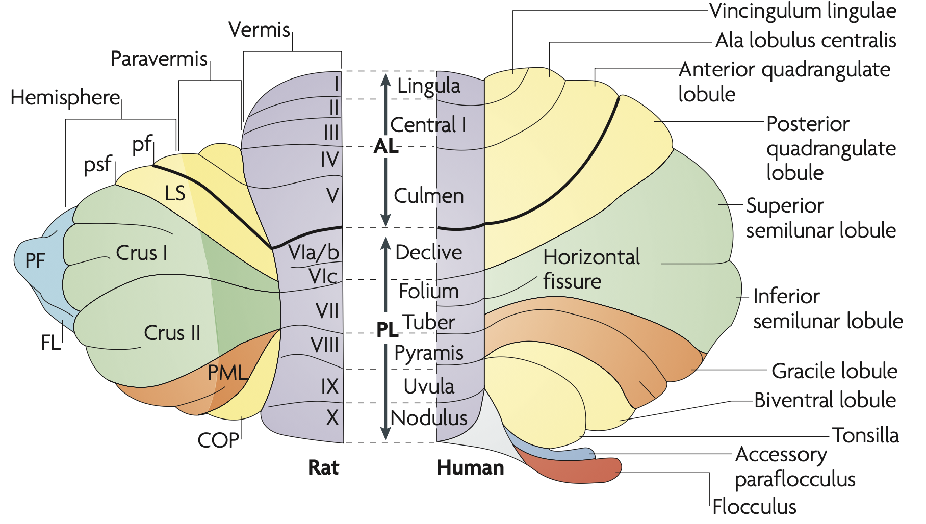
Figure 5: Terminology for the anatomy of the cerebellar cortex in the rat and human. The general evolutionary progression is from the medial vermis outward to the more lateral areas. Although the lobes have all been labeled, they are not very functionally relevant; the most relevant organization is the medial to lateral organization. The appendage-like flocculus (FL) and parafloculus (PF) are specialized areas that process the combination of vestibular, visual, and visuomotor signals. From Apps & Hawkes (2009).
Figure 5 shows the terminology for all of the different regions of the cerebellar cortex, progressing outward along the medial-lateral axis from the original vermis to the paravermis and lateral hemispheres, which are involved in more fine-grained skeletal motor control. There are also unique areas known as the flocculus (FL) and paraflocculus (PF) which process the convergence of vestibular, visual, and visuomotor signals.
The stripe-like structure shown in Figure 4 is due in part to a protein called zebrin II (i.e., aldolase C or ALDOC) that reflects an organizing structure for the long stripes along the length of the cerebellum (Sugihara & Shinoda, 2004, Hawkes & Leclerc, 1987). Although the flattened version of the cerebellum is convenient for visualization, it is actually a massively folded structure, with folds going perpendicular to the long axis of the zebrin stripes.
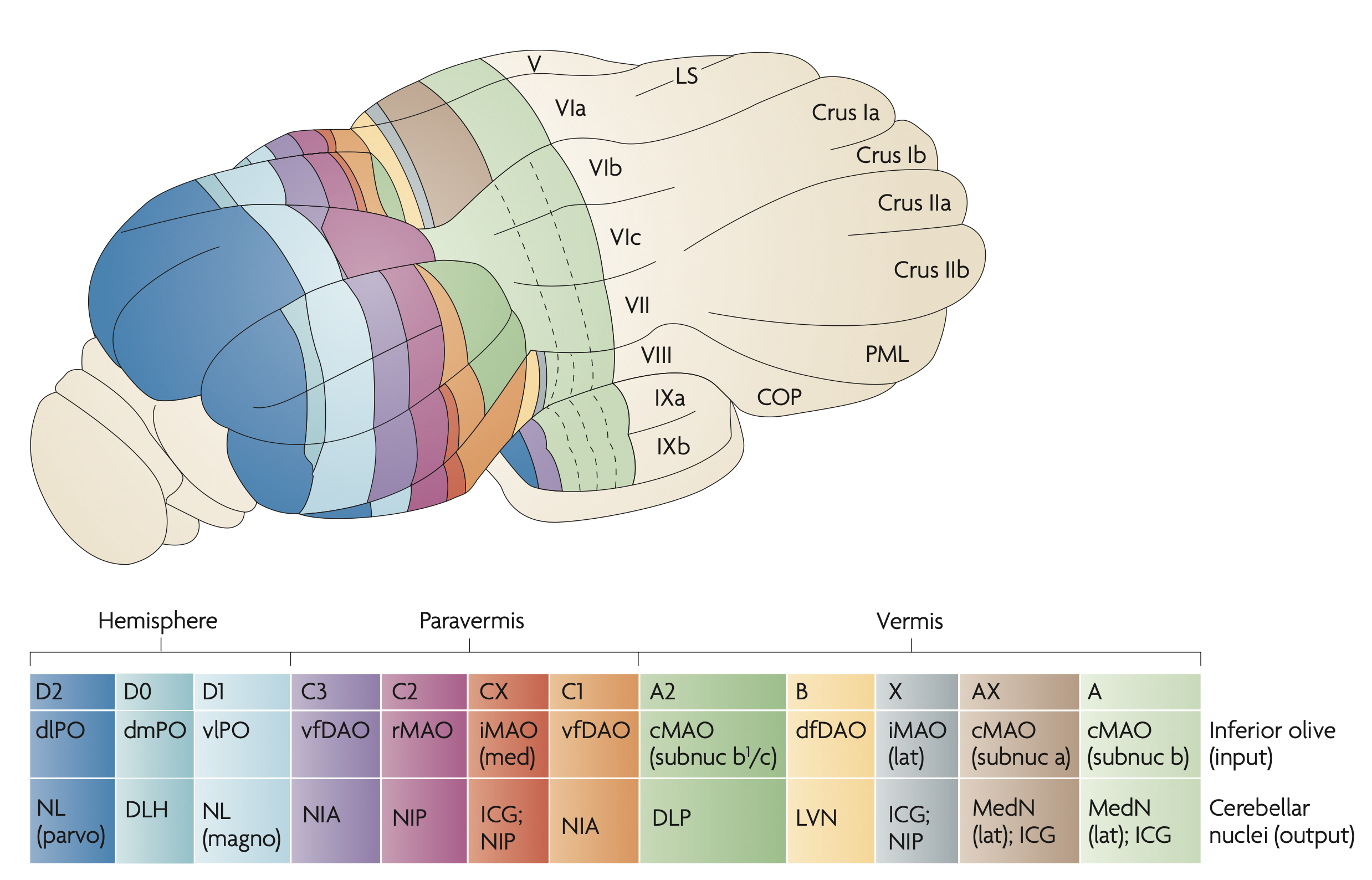
Figure 6: Mapping between the crebellar cortex, the IO (inferior olive), and the different nuclei, The nuclei are: MedN = medial (aka fastigial = FN); LVN = lateral vestibular nucleus; ICG = interstitial cell group; NIP = IPp; nucleus interpositus posterior; DLP = dorsolateral protuberance of the medial nucleus; NIA = IPa = interpositus anterior; NL = lateral nucleus; DLH = dorsolateral hump; From Apps & Hawkes (2009).
Figure 6 shows the relationship between the cerebellar cortex and the different nuclei and areas of the inferior olivary nuclei (IO) (from Apps & Hawkes, 2009). There is different terminology for the nuclei names, with the names in the rodent going from medial (i.e., the fastigial) to the lateral, with the interposed or interpositus and interstitial nuclei in between. The lateral is the most evolutionarily modern nucleus (called the dentate in primates), interconnecting with the lateral hemispheres, which are the areas most enlarged in humans. This is where fine motor control associated with highly dexterous hand movements is supported. The intermediate nuclei are involved in major skeletal muscle control of the limbs, which evolved in land-dwelling animals.
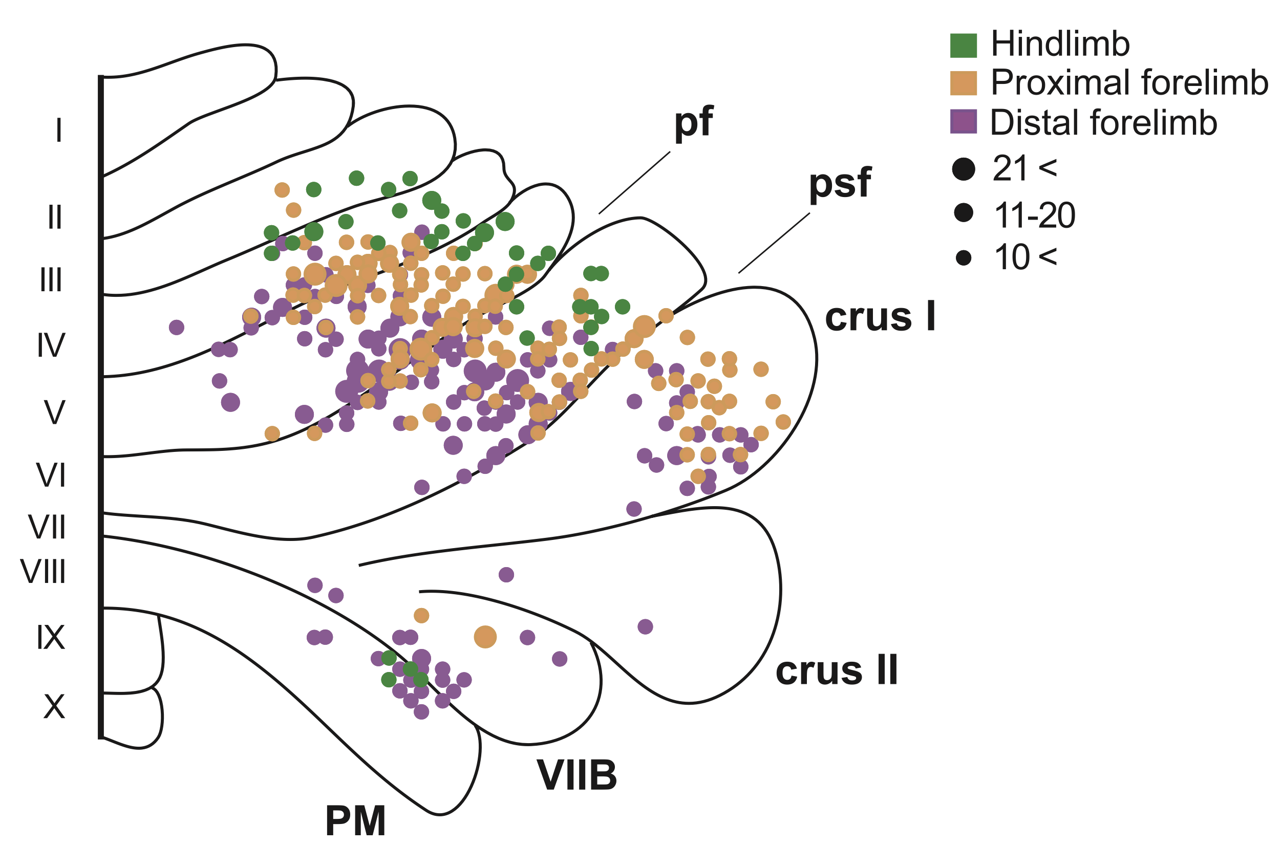
Figure 7: Distributed topographic projections for different skeletal motor areas in the macaque monkey, consistent with the cross product connectivity pattern, where each area is represented in multiple distributed locations. The areas of the lateral cerebellum interconnected with the interposed nucleus, which are associated with the various skeletal muscles, are the only part of the cerebellum with significant topography, where the limb projections are in the same ordering on the medial-lateral axis. From Lu et al., 2007.
Figure 7 shows an example of the somatotopic (topographic) organization of the parts of the cerebellum interconnected with the skeletal muscles, which are those interconnected with the interposed / interpositus (IP) nuclei. Whereas the primary somatosensory (S1) and motor (M1) cortex have a fully somatotopic homunculus (“little man”) organization, the cerebellum retains a more cross-product like structure with more widely distributed projections, but some measure of medial-lateral topography (Lu et al., 2007).
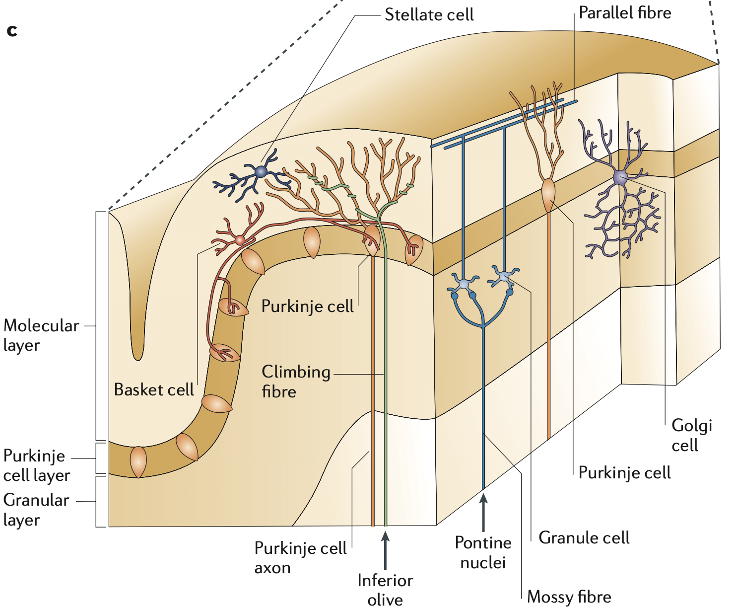
Figure 8: The major anatomical features of the cerebellar cortex. The Purkinje cells (PC) send inhibition to the CN, which is the only output of the cortex. The inputs come via the mossy fibers which then synapse onto tiny granule cells, which send distinctive axons known as parallel fibers to the PCs. Each PC can receive 150,000 or more parallel fiber inputs, via its extremely large dendritic arbor. Each PC receives exactly one climbing fiber from a neuron in the IO, which has been shown to drive synaptic changes (LTD = long term depression) in the parallel fiber synapses. From Ramnani (2006).
Figure Figure 8 from Ramnani (2006) shows the major anatomical features of the cerebellum, many of which are distinctive relative to other brain areas. We will review each of these features in detail in the context of the functional model developed below. The basic circuitry in the cerebellar cortex starts with mossy fiber inputs conveying the different sensory-motor inputs (e.g., from the pontine nuclei) onto millions of tiny granule cells, which then send a huge number of parallel fibers to the only output neurons from the cortex: the Purkinje cells.
The Purkinje cells are tonically active at relatively high rates (~50 hz) and send inhibition to the excitatory cerebellar nuclei output neurons (not shown), which are also similarly tonically active. They receive climbing fiber input from exactly one inferior olivary (IO) neuron, which is known to drive learning in the Purkinje cells, consistent with the original computational models of Marr (1969), Albus (1971), and Ito (1972).
Critically, this circuit supports largely parallel processing across all the loops that are replicated across the entire cerebellum, with very little interaction among the different loops. The granule cell parallel fibers distribute the same sensory input signals efficiently across these loops, but each Purkinje cell output channel is largely independent. This means that other brain areas, principally the basal ganglia and especially the neocortex, are responsible for integration and comparison across these channels in order to support decision making and the hierarchical organization of motor actions. The cerebellum is thus not well-suited for choosing or shaping actions per se, but rather is critical for providing a high-dimensional, coordinated sensory basis for guiding the actions selected elsewhere.
There is one interesting and often neglected direct feedback projection within the cerebellum, which goes from the excitatory output neurons in the cerebellar nuclei back up to the granule cells and golgi cells in the cortex (Houck & Person, 2014; Houck & Person, 2015; Ankri et al., 2015). This can provide a quick broadcast of the current output of the cerebellum, for other cerebellar pathways to learn about. This feedback loop also provides input to the same modules that send Purkinje projections to these cerebellar nuclei neurons, so it provides closed loop as well as open loop feedback.
The starting point for the model we adopt is that the cerebellum provides a stable, effective basis for motor control by learning to predict the sensory effects of motor actions, and to subtract these predicted sensory signals from the ongoing stream of sensory inputs (Fujita, 1982; Dean et al., 2010; Cullen, 2023). This adaptive filtering function can be directly supported by the massive inhibitory projections from the Purkinje cells onto the cerebellar nuclei, which represent the sole output of the cerebellar cortex. The net effect is that any residual sensory signals that have not been canceled therefore represent something beyond the consequences of one’s own actions, and are thus the relevant basis for driving further actions.
The same type of predictive learning mechanism that supports the cancellation of sensory signals generated by one set of motor actions is also useful for driving subsequent motor actions based on the anticipated sensory outcomes of those initial actions. A classic example of this is the widely studied vestibulo-ocular reflex (VOR) where the predicted vestibular signals generated by head movements can then drive corrective eye movements in the opposite direction, to maintain a stable visual stimulus on the retina (Zeeuw et al., 1998; Kimpo et al., 2014). To effectively stabilize the retinal image, it is essential that the eye movements anticipate the effects of the head movements, not just react to them after the fact. This function is consistent with the widely discussed forward model theory of cerebellar function (Miall & Wolpert, 1996; Bastian, 2006; Tanaka et al., 2020).
Many theorists have discussed these functions for the cerebellum over the years, but the precise neural mechanisms by which they are accomplished have remained somewhat underspecified in a few important respects. Furthermore, many other functional frameworks have been proposed that are either more generic (e.g., basic error-driven learning as in the Marr-Albus-Ito models, or feedforward motor control based on the widely studied delayed eyeblink conditioning paradigm; Thompson, 1986; Raymond et al., 1996; Ohyama et al., 2003; Nguyen & Person, 2025) or more complex (e.g., Kawato, 1999; Llinás, 2009).
Further complicating matters is the relationship between processing and learning in the cerebellar cortex and that taking place in the cerebellar nuclei, which have been comparatively neglected in the literature (Kebschull et al., 2024; Herzfeld et al., 2020). There has long been evidence that the cerebellar nuclei are the final long-term locus of consolidated cerebellar learning, with the cerebellar cortex therefore playing a more transient role (Kassardjian et al., 2005; Shutoh et al., 2006; Kimpo et al., 2014; Raymond & Medina, 2018). Thus, a complete account of the underlying neural mechanisms must explain how the circuitry within the cerebellar nuclei can independently support the full predictive learning functionality, and how learning within the nuclei relates to that taking place in the cerebellar cortex.
The cerebellar cortex certainly provides a much higher-resolution representation of the sensory-motor space, due to the huge number of granule cells, but also due to a large expansion factor in the number of Purkinje cells (PC) relative to the number of neurons in the cerebellar nuclei (CN). Bengtsson et al. (2011) estimate that between 600-1,200 PC’s converge onto an individual CN output neuron, with estimates of ~8,500 total CN excitatory output neurons per hemisphere in a mouse (Heckroth, 1994), versus nearly 200,000 PC’s (Woodruff-Pak, 2006).
The Nuclear model described below focuses first on the computations in the cerebellar nuclei (CN), showing how connectivity and plasticity among the CN neurons can directly support both adaptive filtering and forward model functionality, building off of a predictive learning mechanism driven by one specific type of inhibitory CN neuron. We hypothesize that these inhibitory CN neurons learn to predict and anticipate a specific sensory input channel, and they project into the inferior olive (IO), which also receives a precisely paired excitatory projection from the same sensory input. This provides an explicit neural mechanism by which the IO can directly compute the sensory prediction error as the difference between these paired excitatory and inhibitory inputs.
The paired IO inputs are typically integrated within an elaborate glomerulus structure on the IO dendrites, which has complex neural properties that appear ideally configured to perform a temporally offset error computation (De Zeeuw et al., 1998; Loyola et al., 2023). The IO neurons project this critical error signal to both the CN and the Purkinje cells in the cerebellar cortex, driving learning throughout the cerebellum.
Although the properties of this error signal have been widely discussed as strangely impoverished (Najafi & Medina, 2013; Zang & De Schutter, 2019), we argue below that it actually makes good sense for driving precisely timed learning, at the cost of communicating more graded information, which would compromise the temporal precision.
Thus, the neural mechanisms in the Nuclear model provide a complete account of coordinated learning within the cerebellar nuclei and the much larger cerebellar cortex. The cortical learning mechanisms are faster than those in the nucleus, and they are also subject to decay (Herzfeld et al., 2020; Kimpo et al., 2014; Kassardjian et al., 2005). By contrast, the nuclear learning is slow and persistent. In short, the CN slowly learns the enduring rules, while the cerebellar cortex can quickly learn the exceptions, just as in the complementary learning systems framework for the hippocampus vs neocortex (McClelland et al., 1995; O’Reilly & Rudy, 2001; Norman & O’Reilly, 2003). If the exceptions persist, and especially during initial developmental learning, then they are consolidated into the CN.
After presenting the Nuclear model, we compare it against other models in the literature, with the goal of understanding how the specific contributions of the cerebellum fit into the broader motor system, to support flexible, adaptive, and capable motor behavior.
The Nuclear model
Adaptive filtering
The predictive cancellation function performed by the cerebellum is known as an adaptive filter (Fujita, 1982; Widrow & Stearns, 1985), which is a form of predictive learning that filters out what is expected or predicted based on other sensory signals, so that the residual signal represents a “pure” sensory signal that clearly represents anything that is otherwise unexpected.
When you move any part of your body, especially the head or eyes, you create massive changes in incoming signals across all of your senses (i.e., sensory reafferance). If you did not somehow subtract these self-motion sensory signals in your perceptual system, you would never be able to act sensibly in the world (Cullen, 2023). Everything would be in a constant state of turbulent motion, and you would never be able to coordinate a proper motor action based on all of these moving targets.
The evolutionary history of the cerebellum is consistent with this adaptive filter function, based on proto-cerebellar circuits and cerebellar analogs in other animals (Bodznick et al., 1999; Montgomery & Perks, 2019; Bell et al., 2008; Cisek, 2021). The most ancient part of the mammalian cerebellum is essentially an integral component of the primary brainstem vestibular sensory nucleus, where it learns to subtract the effects of self-generated motion signals at all levels of the body, to produce a pure vestibular signal that represents any remaining discrepancy from what would be expected based on the self-motion generated signals.
This pure vestibular signal is then something that can be acted upon effectively. If there is an additional sense of motion after all of your own actions have been accounted for, you know that you are slipping or falling or being pushed around in some way. Furthermore, this pure signal tells you exactly which way you should act in order to counteract the residual motion. When combined with other sensory inputs from vision, touch and proprioception, you can also figure out what the likely cause of the disturbance is, and thus condition your motor response accordingly.
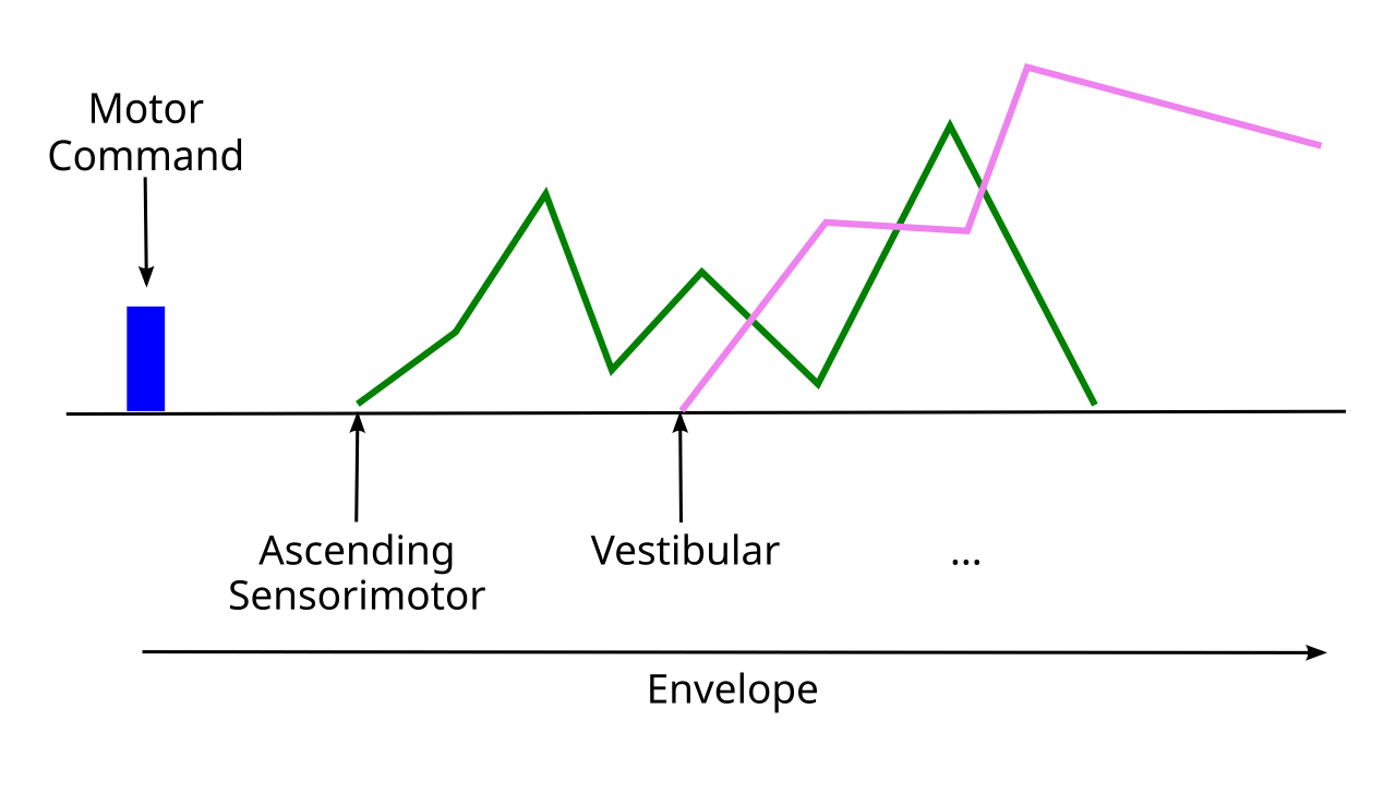
Figure 9: The temporal envelope surrounding a descending motor command, with temporal delays offsetting from the start of the command. A cascade of ascending sensorimotor signals from the spinal cord tracks the unfolding of the motor action (reflections of actual muscle signals, proprioceptive muscle spindle fiber signals, etc). The vestibular signal reflects the head and body motion in terms of velocity and acceleration in three dimensions.
As this example makes clear, the adaptive filtering by the cerebellum provides an essential basis to enable sensible motor actions to be generated. A major challenge that the cerebellum must overcome is managing the many different temporal delays associated with the effects of motor actions and the sensory transduction thereof. When you decide to move your head, the efferent copy of that descending motor command provides the earliest signal about what is going to happen, setting off a complex cascade of subsequent signals that all need to be accounted for (Figure 9).
Your spinal cord and skeletal muscles do a lot of further work to actually implement the motor command in an efficient way, so you need to also get the actual ascending motor signals back up from the lower-level spinal motor pathways and the associated proprioceptive signals about the current levels of muscle stretching from the muscle spindle fibers. These signals unfold over time as the motor action takes place, even if the descending motor command was relatively ballistic.
Then, as your head starts to move, you will start getting signals from your vestibular system reflecting the motion. The specific spatiotemporal pattern of these signals will depend on the exact motor parameters, so this is a further challenging prediction problem. As shown in Figure 9, the adaptive filtering job performed by the cerebellum can be organized around the entire temporal envelope defined by the initial descending motor command.
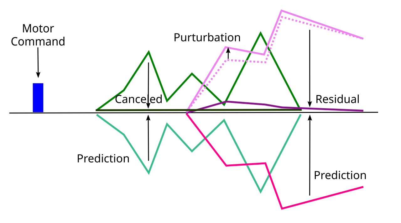
Figure 10: The prediction of the sensory consequences of a motor action can be subtracted from the original sensory signal to cancel the sensory signal. If there is an external perturbation of the sensory signal that does not fit the prediction, then there is a residual signal that clearly reveals the exact parameters of the perturbation, untangled from the sensory signals generated by the motor command.
The core computation performed by the adaptive filter is to subtract the learned prediction of the sensory consequences of a motor action from the actual sensory inputs (Figure 10). If the actual sensory inputs match the learned prediction, then the result is cancellation of the sensory signal. However, if there is a perturbation of the sensory signal from something else unexpectedly going on (a slip, a push, etc), then this will show up clearly as a residual signal that has not been canceled by the prediction.
Forward models
In addition to the inhibitory cancellation process, the learned sensory prediction signals themselves are directly valuable in supporting motor control, because they provide anticipatory information about the sensory outcomes of motor actions. This anticipatory prediction of sensory outcomes for use in motor control is known as a forward model (Jordan & Rumelhart, 1992; Shadmehr et al., 2010), which has long been hypothesized as a critical aspect of cerebellar processing (Miall & Wolpert, 1996; Bastian, 2006; Tanaka et al., 2020).
The distinction between a forward model and the adaptive filter is often not clear in the literature, because both involve the same core computation of predictive learning of sensory signals. In the terminology adopted here, we define a forward model as using these predictive signals to directly drive motor actions based on anticipated states, whereas an adaptive filter removes these predicted signals from the sensory states.
The adaptive filter requires increased inhibition in proportion to the predicted sensory signal to drive the cancellation, whereas the forward model signal should go in the opposite direction, to be proportional to the sensory prediction. Both should be driven by a predictive learning mechanism triggered by the efferent copy of a motor action, so that they are “about” the action, and not just always learning to cancel everything.
One clear example of these two functions operating on the same vestibular signal is the adaptive filtering of head motion to subtract from the vestibular signal (Cullen, 2023), versus the vestibulo-ocular reflex (VOR) where eye movements are generated to correct for head motion, to keep a visual stimulus stable on the retina (Zeeuw et al., 1998; Kimpo et al., 2014; see VOR simulation). The VOR requires ocular motor actions to be generated in anticipation of the effects of head motion actions, which requires the forward model predictions.
To be able to anticipate sensory signals and respond as quickly as possible, the cerebellar circuits all operate in parallel. Thus, the adaptive filtering and forward model functions, though logically related, operate via parallel loops. It would be too slow for the VOR ocular actions to be generated by sequentially feeding a signal from the adaptive filtering circuit back into another cerebellar circuit for the VOR.
The cerebellum is organized to support the specific demands of adaptive filtering and forward models, in parallel. The cerebellar nuclei directly implement the common predictive learning aspect, and also send a prediction error signal to the cerebellar cortex via their inputs into the inferior olivary nuclei. Critically, this IO error signal can drive different directions of plasticity in different microzones, known as upbound (zebrin II positive) and downbound (zebrin II negative; De Zeeuw, 2021). These different microzones can then support the adaptive filtering and forward model functions, respectively.
Given the considerable evidence that cerebellar learning is consolidated into the circuits of the cerebellar nuclei (Kassardjian et al., 2005; Shutoh et al., 2006; Kimpo et al., 2014; Raymond & Medina, 2018; Herzfeld et al., 2020; Kebschull et al., 2024), these circuits need to be able to directly implement the adaptive filtering and forward model functionality. Understanding how this could work provides clear, novel functional explanations for many features of the cerebellar system.
Nuclear circuitry
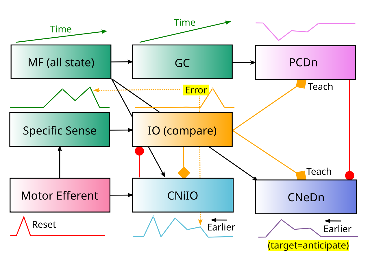
Figure 11: Cerebellar components that implement the Nuclear model of forward model functionality in downbound (Dn) microzones. The target output for the CNeDn excitatory CN (cerebellar nucleus) output is to anticipate the sensory consequences of motor actions for a given Specific Sense input, as early as possible, as indicated by the temporal activity trace schematized under each neuron type. The Motor Efferent copy signal provides the initial predictive trigger, so that the prediction is about a specific action. This trigger arrives before (e.g., 50-100 ms) the sensory effects it causes, so the IO (inferior olive) computes an offset comparison of later sensory inputs vs. the anticipatory CNiIO inhibitory IO prediction, with the motor trigger resetting IO dynamics. When excitatory sensory input arrives without a prior CNiIO inhibitory prediction, the IO fires a “learn now” teaching spike, to correct the error. IO spikes drive learning in the CNeDn neurons to modify inputs from MF (mossy fiber) time-varying state inputs, which can then drive earlier anticipatory firing of these CNeDn neurons. CNeDn also contributes back to the MF inputs for rapid sharing with other cerebellar pathways. The PCDn (Purkinje cell) neurons use high-dimensional, rapidly time-varying granule cell (GC) representations of MF state to generate more precise, rapidly adapting predictions to disinhibit the CNeDn anticipatory output, trained by IO climbing fiber complex spikes that result in LTD.
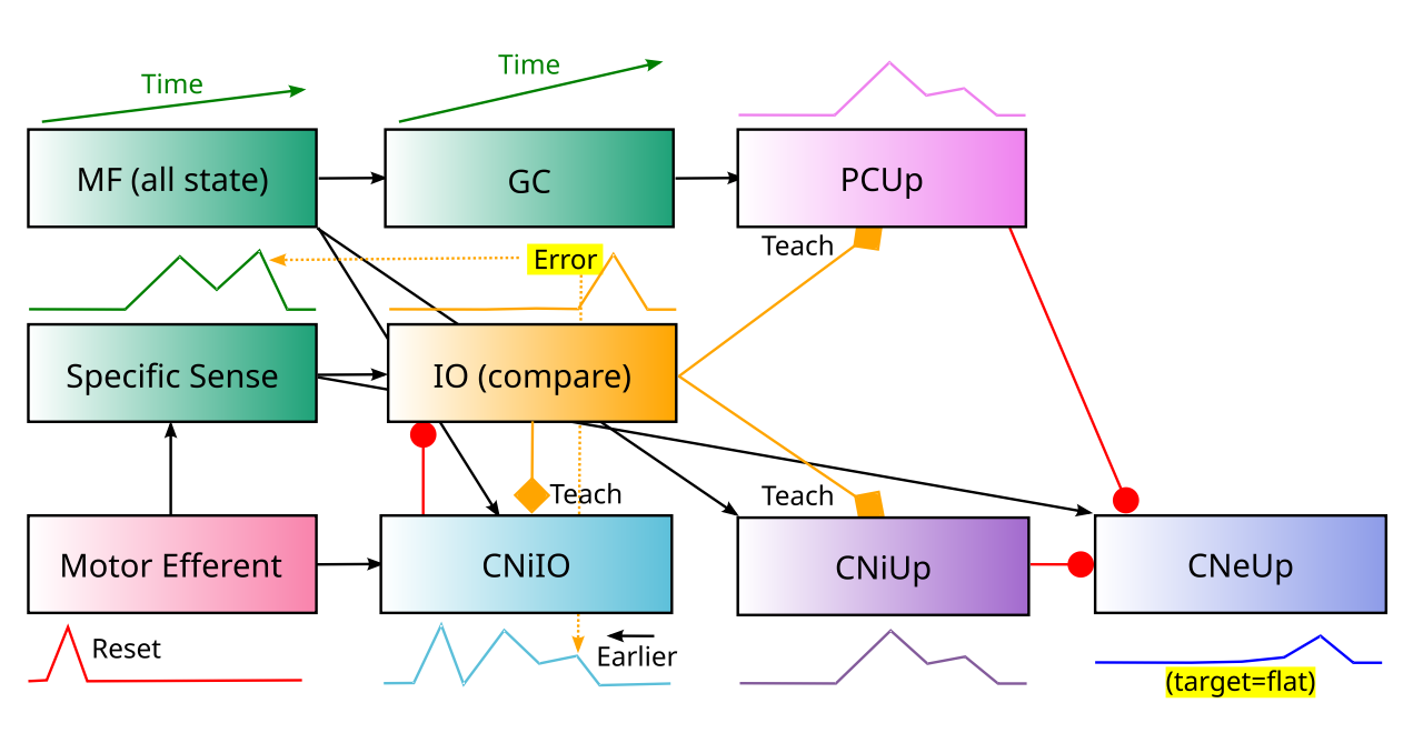
Figure 12: The Nuclear model of adaptive filtering in the upbound (Up) microzones. See prior figure for shared elements, with key differences noted here. The target output in the CNeUp excitatory CN output neurons is flat tonic activity, with deviations reflecting unexpected signals from the Specific Sense input. Unlike the forward model case, the local inhibitory neurons (CNiUp) must learn to inhibit the CNeUP output at the time when subsequent sensory input actually arrives, which is generally delayed by up to 50-100ms from the motor efferent trigger and the anticipatory CNiIO activity. Therefore, the CNiUp neurons learn from the IO error signals to modify their MF inputs to cancel the predicted sensory input that is directly exciting CNeUp. The PCUp Purkinje cell neurons learn to increase firing (LTP) in response to IO climbing fiber complex spikes, to increase inhibitory cancellation of the CNeUp output.
Figure 11 shows the key elements of the cerebellar circuitry that can implement the forward model process according to the Nuclear model, while Figure 12 shows the adaptive filtering case. Even though each major element in this model has been documented and discussed in the literature, the specific details have not otherwise all been combined into one coherent framework. The points of comparison relative to Standard models are discussed below, after providing an overview of the Nuclear framework.
Each specific sensory channel (e.g., one particular canal in the vestibular system) has an anatomically organized collection of coordinated cerebellar nucleus (CN), inferior olive (IO), and cerebellar cortex pathways that specifically learns to predict it, based on the microzone organization (Oscarsson, 1979; Sugihara & Shinoda, 2004; Hawkes & Leclerc, 1987; De Zeeuw, 2021). A key additional hypothesis is that the same specific sensory inputs that project to the IO for a given microzone also project to the corresponding CN neurons for the upbound microzones, where they are necessary for driving output activity reflecting the sensory signal that is not otherwise expected from motor actions.
The IO projections provide a “learn now” teaching signal to both the CN neurons (Want & Voogd, 1987; De Zeeuw et al., 1997; Ruigrok & Voogd, 2000; Baumel et al., 2009; Rowland & Jaeger, 2005) and the PC neurons via the well-documented climbing fiber and complex spike mechanisms. This IO signal reflects a discrepancy between the CNiIO inhibitory prediction and the specific sensory input. Because the CNiIO prediction is triggered by the early motor efferent signal, it arises roughly 50-100ms in advance of the consequent sensory signals, so the IO requires specialized time-offset mechanisms to allow the earlier prediction to be compared against the later sensory input.
The forward model case in downbound microzones operates entirely on the early timescale, to provide maximally anticipatory predictions of the effects of motor actions. By contrast, the adaptive filtering case in upbound microzones needs to drive cancelling inhibition synchronized with the later sensory input signals. This key distinction is consistent with latency differences between upbound and downbound pathways (ten Brinke et al., 2015), and it requires different connectivity and learning logic within the CN. Data on this level of microzone-specific connectivity does not yet exist (with a few notable exceptions discussed below), so the Nuclear model therefore generates several testable predictions.
The CN circuitry described first provides a complete implementation of the adaptive filtering and forward model functionality, consistent with the data showing that this is the locus of consolidated, long-term cerebellar learning (Kassardjian et al., 2005; Shutoh et al., 2006; Herzfeld et al., 2020; Kimpo et al., 2014). There is evidence for plasticity in the CN neurons in the intact brain (Medina & Lisberger, 2008; Kimpo et al., 2014; Yang & Lisberger, 2014; Herzfeld et al., 2020) along with direct tests of CN plasticity in slices (Aizenman et al., 2000; Pugh & Raman, 2008; Pugh & Raman, 2006; Zhang & Linden, 2006; McElvain et al., 2010).
The CNiIO neurons also drive learning in the cerebellar cortex via their impact on the IO climbing fiber complex spikes. The cortex can provide a more rapid and temporally precise version of the forward model and adaptive filtering functions, across the upbound and downbound microzones. After presenting the specific components of the Nuclear model, a more detailed consideration of the learning functions for each element of the system is presented, including the actual equations used in the model.
Specific sensory and motor inputs
A critical element of the Nuclear model is that each microzone is organized around the processing of a specific sensory input signal, for example a vestibular canal, which is either anticipated (in the forward model case) or canceled (in the adaptive filtering case). Furthermore, these functions are contingent on a specific action (e.g., head rotation), which occurs significantly prior to its sensory sequelae.
Table 1: Anatomical inputs to CN, Cerebellar Cortex (Kebschull et al., 2024) (Med = medial, PIN = posterior interposed, AIN = anterior interposed, Lat = lateral)
| Input | Modality | Med | PIN | AIN | Lat | Cortex |
|---|---|---|---|---|---|---|
| Spinal cord | ||||||
| Cervical | Neck, arm, hand | ++ | ++ | ++ | – | +++ |
| Thoracic | Trunk | ++ | ++ | ++ | – | +++ |
| Upper lumbar | Hip, legs | ++ | ++ | ++ | + | +++ |
| Sacral, lower lumbar | Foot, toes | ++ | + | +++ | – | +++ |
| Column of Clarke | Spindle fibers | + | – | + | – | +++ |
| Medulla oblongata | ||||||
| Cuneate, gracile | Somatosensory | – | – | – | – | ++ |
| Spinal trigeminal | Face somatosensory | ++ | ++ | ++ | ++ | +++ |
| Vestibular ganglion | Primary vestibular | – | – | – | – | ++ |
| Medial Vestibular (MVN) | Secondary vestibular | ++ | – | – | – | ++ |
| Reticular formation | ||||||
| Paramedian (PPRF) | Gaze (SC, vestibular) | ++ | ++ | ++ | ++ | +++ |
| Giganto, magnocellular | Posture, trunk, tone | ++ | ++ | ++ | ++ | ++ |
| Lateral (LRN) | Reflexes | ++ | ++ | +++ | + | +++ |
| Red nucleus (magnocellular) | Gait, grasping | – | + | ++ | + | – |
The specific sensory input signals originate from various spinal and midbrain nuclei, including the vestibular nuclei, trigeminal nuclei, dorsal column nuclei, medulla oblongata, and reticular nuclei, as shown in Table 1 (adapted from Kebschull et al., 2024, which is not exhaustive). These are generally consistent with the inputs to the IO (Swenson & Castro, 1983; Brown et al., 1977), although the IO receives from the cuneate and gracile nuclei (Berkley & Hand, 1978), whereas the CN do not (Dietrichs & Walberg, 1987; Quy et al., 2011).
The specific sensory inputs (and the MDJ pathway as discussed below) are particularly critical for the upbound adaptive filtering function in the CN, as shown in Figure 12. Specifically, the sensory input must drive the CNeUp output neurons so that they correctly reflect any unpredicted sensory signals that are not canceled by the CNiUp and PCUp inhibition.
There are several lines of evidence that are consistent with the upbound areas receiving specific sensory inputs. At a broad level, while many cerebellar inputs that form the mossy fiber pathway in the cerebellar cortex also project to the CN, there are also exceptions (Table 1), which are generally consistent with the CN projections being more from lower-level “basic” sensory pathways, versus higher-level integrated areas. For example, only about 1/5th of pontine nuclei neurons (which convey neocortical inputs) had branches into the CN (Shinoda et al., 1992), whereas neurons in the lower-level lateral reticular nucleus (LRN) all had CN projections (Wu et al., 1999).
Furthermore, the CN projections are generally more focal than those to the cerebellar cortex (Sugihara, 2011; Ando et al., 2020; Shinoda et al., 1992; Wu et al., 1999). In addition, the CN areas that project strongly to the MDJ are zebrin II positive, i.e., upbound (Sugihara, 2011; see also Wang et al., 2022).
Table 2: Functional groups (Sugihara, 2011). (v = ventral, d = dorsal, m = medial, l = lateral, c = caudal, r = rostral; FN = fastigial, ICG = interstitial cell group, PIN = posterior interposed, DN = dentate, DLP = dorsolateral protuberance, LVN = lateral vestibular nucleus, DMC = dorsomedial crest, AIN = anterior interposed; MD = Mesodiencephalic)
| Group | Zebrin II | Inputs | CN | Cortex | Zone | IO |
|---|---|---|---|---|---|---|
| I | + Up | Mesodiencephalic | FN v m | 1+//1+ | A | c-MAO c |
| FN v l | 2+//3+ m | AX | ||||
| ICG v | 2+//3+ l | AX | c-MAO r | |||
| PIN dl | 4+//5+ | C2 | r-MAO | |||
| DN c | 5+//6+ | D1 | v-PO | |||
| DN rl | 6+//7+ | D2 | d-PO | |||
| IIa | + Up | Vestibular | FN vc m | a+//2+ m | A | Beta c |
| FN vc l | a+//2+ l | A | Beta c-r | |||
| PIN v | 2b+//4+ | X-CX | DMCC, DM c, v-PO cm | |||
| IIb | + Up | Collicular | FN mid-c m | a+//2+ | Lat A | c-MAO m |
| FN mid-c dl | c+//4b+ | Lat A | c-MAO l c | |||
| FN mid-c l | d+//5a+ | X-CX | c-MAO r | |||
| III | – Dn | Soma, Vest, MD | FN rd m | 1-m//1- | A | c-MAO l c |
| FN rd l | 1-l/a-//2- | A1 | c-MAO int | |||
| ICG d | 2a-//3- | X | c-MAO r | |||
| DLP r | 2b-//4a- | Lat A | c-MAO m cl | |||
| DLP d | c-//4b- | Lat A | c-MAO cm | |||
| DLP c | d-//5a- | Lat A | c-MAO int | |||
| PIN dm | 3b-//e2- | CX | c-MAO r | |||
| IV | – Dn | Somatosensory | LVN, AICG | 2-//4- | B | d-DAO m c |
| LVN, AICG | b+//f+ | B | d-DAO l pole | |||
| DMC (cm AIN) | 3+//el+ | C1 | v-DAO cl | |||
| AIN rm | b-//f- | C1 | v-DAO l | |||
| AIN c | 3-//el- | C1 | v-DAO c | |||
| AIN l | 4-//5- | C3 | v-DAO rm | |||
| V | + Up | Visual | v-DN, d-Y | floc, nod | D0 | DC, VLO |
Table 2 adapted from Sugihara (2011) (see Figure 5 and Figure 6 for locations, in the rat) provides an attempt to organize the different upbound and downbound microzones into groups based on their inputs. From this, it seems that the downbound (forward model) areas are more diverse in their inputs, and overall much more focused on somatosensory inputs. By contrast, upbound (adaptive filtering) is focused more on vestibular and visual, auditory localization signals (collicular), while somatosensory signals are not significantly filtered.
These differences in sensory inputs make sense in terms of the sensory signals that are used to drive motor actions, which require upbound adaptive filtering so that the sensory signal properly compensates for other actions. These sensory inputs are largely visual and auditory localization inputs, which provide a distal guide to where you want to reach or navigate to. Indeed, a range of evidence shows that most motor areas in cortex use visually grounded spatial reference frames for planning (e.g., Batista et al., 1999). Because these sensory inputs (eyes and ears) are located on the head, they also require knowing about the orientation of the head, as provided by vestibular inputs.
By contrast, somatosensory inputs provide feedback signals that result from motor actions, but do not represent the target or goal of actions. Therefore, they are important to anticipate for coordinating actions in the downbound forward model areas, but not for adaptive filtering.
The connectivity with the mesodiencephalic system listed in Table 2 is based on the projections from the mesodiencephalic junction (MDJ) into the relevant areas of IO, which in turn are known to be associated with corresponding projections from CN output neurons into the same MDJ areas (De Zeeuw et al., 1998; Wang et al., 2022; De Zeeuw & Ruigrok, 1994; De Zeeuw et al., 1998; Rutherford et al., 1989). However, this does not mean that the MDJ areas also send specific sensory inputs into these CN areas. Instead, the CN areas receive from the other areas that the MDJ neurons also receive from, which are generally quite widespread (Khalil et al., 2022; Onodera & Hicks, 1995).
For example, one of the MDJ nuclei, the nucleus of Darkschewitsch, serves as an integrator and coordinator of many different occulomotor-related areas, including eye-related motor cortical areas, SNr and GPe (basal ganglia output), the superior colliculus, and other mesodiencephalic structures such as the zona incerta (ZI) and posterior pretectal nucleus (Onodera & Hicks, 1995). These other inputs can be either excitatory or inhibitory. Perhaps the IO signal is driven by the more integrative MDJ neurons because that provides a more behaviorally relevant level of error signal, whereas the CN adaptive filtering is more effective to apply to more elemental sensory signals.
Other MDJ nuclei include the red nucleus, nucleus interstitialis of Cajal, nucleus of Bechterew, tegmental field of Forel, zona incerta, subparafascicularis nucleus, and the prerubral reticular formation (De Zeeuw et al., 1998). The magnocellular red nucleus (mRN) is interesting in Table 1, because it shows the opposite pattern of most other inputs, projecting to (and receiving from) the interposed CN but not the cerebellar cortex (Beitzel et al., 2017). It is critical to distinguish the mRN pathway, which drives the rubrospinal motor projections into the spinal cord (Basile et al., 2021), from the parvocellular pRN, which is a component of the MDJ areas, and interconnects with the IO and corresponding CN areas as noted above.
The distinction between these two aspects of the red nucleus is anatomically clear in primates, but not in other species, where they are more intermixed (Liang et al., 2012; Basile et al., 2021). Whereas the function of the pRN remains mysterious in the literature, the mRN has been clearly demonstrated to provide coordinated motor control of distal limbs for quadrupedal locomotion and grasping. It is likely that discussions of the red nucleus in the literature often confuse the functions of these distinct pathways (e.g., Wang et al., 2022).
CNiIO prediction learning
The inhibitory (GABAergic) CNiIO neurons learn to predict the specific sensory channel activity, based on learning driven by the excitatory sensory inputs. The IO “learn now” input can trigger learning in the mossy fiber (MF) inputs, to support error-driven learning that predicts the sensory inputs. The sample of time-varying inputs in the MF allows the CNiIO neurons to generate a prediction of the time-varying specific sensory input.
Because synaptic plasticity mechanisms reflect the integration of earlier synaptic input activity, these MF-to-CNiIO connections learn to anticipate the subsequent sensory signal (Raymond & Lisberger, 1998; Wang et al., 2000). This anticipation is consistent with an initial activation by the motor efferent signal that precedes the sensory sequelae of the action, so the CNiIO predictive activity remains anticipatory, which is necessary for the forward model functionality.
The CNiIO neurons are significantly less active at rest than the other CN neuron types (Kebschull et al., 2024; Uusisaari & Knöpfel, 2011; Özcan et al., 2020), consistent with the idea that they are predicting the specific sensory input signal and not exerting tonic inhibitory control over sensory and motor systems, as the principal output CN neurons are thought to do.
The learning in CNiIO neurons has been hypothesized to be influenced by PC input (Pugh & Raman, 2008; Yang & Lisberger, 2014; Herzfeld et al., 2020). However, unlike the CNe neurons, it is important that the CNiIO neurons not receive too much inhibition from the PC neurons, as this would then reduce their prediction signal, creating more of a prediction error. This requirement is consistent with evidence showing that PC inhibitory inputs onto CNiIO neurons are relatively weak and synapse on distal dendrites, while PC to CNe projections are strong and synapse near the soma (Uusisaari & Knöpfel, 2008; Uusisaari & Knöpfel, 2011; De Zeeuw & Berrebi, 1995).
IO temporally offset error signal
The CNiIO inhibitory neurons send their learned inhibitory projections into the inferior olive (IO) neurons in the associated microzone. These inhibitory inputs converge with excitatory input from the specific sensory input, often in glomeruli, which are relatively large anatomical structures on IO dendrites, where multiple axons and dendrites converge. By detecting the temporally offset correspondence of excitatory and inhibitory inputs within a narrow time window, the IO glomerulus can compute the prediction error (Ito, 2013; De Zeeuw et al., 1998; De Gruijl et al., 2013).
In rats, each IO neuron has about 50 such glomeruli, each of which receives 2 excitatory and 2 inhibitory inputs (De Zeeuw et al., 1998). Each glomerulus connects about 5-6 different nearby IO neurons together, with gap junctions that cause them to be synchronized. Thus, there are about 5-6 IO neurons processing each specific sensory input projection, with likely redundancy in sampling the same specific sensory channel across cerebellar microzones. For example, about 30 Purkinje cell axons converge onto a single CNe neuron, and one Purkinje cell can innervate at least 20-50 CN cells (Aizenman et al., 2000).
The error detection logic in the IO glomeruli operates by virtue of inhibitory inputs resetting intrinsic dendritic conductances, such that excitatory inputs are ineffective within a ~50-100 ms window, but are strongly amplified outside of that window (Loyola et al., 2023). Thus, even a few spikes outside of the window can drive supra-threshold burst activity in the IO neuron. This time window offset within the IO is essential to allow the CNiIO neurons to exhibit an anticipatory predictive signal. If inhibitory input occurs without a corresponding excitatory input, the neuron remains inhibited for a corresponding time window. The potential mechanisms for correcting excessive predictions in such a case are discussed below.
The complex combination of intrinsic currents in the IO neuron result in a baseline activity level of about 1 Hz, with the relative balance and timing of inhibitory and excitatory inputs determining the probability, timing, and intensity of its spiking (Titley et al., 2019; Zang & De Schutter, 2019; Guo & Uusisaari, 2025; Yang & Lisberger, 2014). At a minimum, these IO spikes can provide a coarse signal that activates plasticity in both the CN and PC neurons that it projects to, with the quantitative details of the learning determined by other inputs.
Furthermore, there is at least some evidence that the IO signal can provide a meaningful bidirectional error signal for learning in PC neurons, with the consistent presence or absence of IO firing associated with sensory inputs driving opposite directions of synaptic plasticity, at least in some well-documented cases (Ohmae & Medina, 2015; Yang & Lisberger, 2014).
It is also likely that the gap-junction driven synchronization of multiple IO neurons is an essential component of the error signal efficacy, with synchronized activity or inhibition across a population providing more of a graded, bidirectional error signal (Hoang et al., 2023; Giessen et al., 2008). This gap junction connectivity could provide a mechanism for organizing a group of related neurons around a common initial motor action signal that defines the temporal envelope (Figure 9).
There is direct evidence showing that the CNiIO projection is responsible for reducing IO firing and complex spikes in PC neurons as learning proceeds (Kim et al., 1998; Ito, 2001). Furthermore, this pathway was shown to be responsible for a blocking effect in conditioned learning, which is a signature of the elimination of prediction error signals in a delta-rule like mechanism (Fanselow, 1998; Kim et al., 1998).
Relative to the well-documented effects of IO spiking on PC neurons (Ito, 2001; Coesmans et al., 2004; Gao et al., 2012; Silva et al., 2024), there is comparatively little known about how the IO inputs could drive learning on CN neurons. The relevant studies of CN plasticity in slices (Aizenman et al., 2000; Pugh & Raman, 2008; Pugh & Raman, 2006; Zhang & Linden, 2006; McElvain et al., 2010) focused on post-inhibitory rebound bursting that is not robustly present in awake behaving animals (i.e., in activo; Hoebeek et al., 2010; Bengtsson et al., 2011). For example, Bengtsson et al. (2011) recorded spontaneous IO background spiking impacts on CN neurons, which generated “giant” IPSPs, but found no sign of rebound firing.
Furthermore, the existing studies are likely to sample the large, tonically active CNe neurons, rather than the smaller, more quiescent CNi neurons that are the critical sites of plasticity according to the Nuclear model. These CNi neurons receive only distal, weak PC input, so they are not as strongly inhibited, and are therefore even less likely to exhibit strong rebound activity.
Despite the limited data, there are multiple lines of evidence that IO activity has significant impacts on CN neurons (Hoebeek et al., 2010; Blenkinsop & Lang, 2011; Baumel et al., 2009; Rowland & Jaeger, 2005), and at least one report of evidence of short-term plasticity effects from IO inputs onto CN neurons in awake animals (Gruart et al., 1994).
CNeDn forward model anticipatory firing
The forward model functionality happens in the the downbound microzones (Figure 11), where the sensory prediction error in the IO neurons drives learning on the CNeDn CN excitatory output neurons. Because the CNeDn neurons do not need to precisely cancel the later-arriving specific sensory input, they can fire as early as possible, to give more time for subsequent motor actions to be shaped in anticipation of the predicted effects of earlier actions. This is the primary hypothesized computational benefit of the downbound forward-model pathway.
The IO climbing fibers project to the CN, and can drive learning from the MF inputs to the CNeDn neurons, producing the requisite excitatory activity, which can anticipate due to the interaction of the synaptic integration process and learning as discussed earlier (Raymond & Lisberger, 1998; Wang et al., 2000). This is consistent with various theories (Pugh & Raman, 2008; Yang & Lisberger, 2014; Herzfeld et al., 2020).
Interestingly, there are also some direct projections from CNiIO neurons into the pontine nuclei and midbrain / brainstem motor areas (Judd et al., 2021), which could thereby send inhibitory anticipatory signals, to complement the excitatory CNeDn ones.
CNeUp adaptive filtering
Although the initial evolutionary function of the cerebellum is thought to be adaptive filtering, it requires some additional mechanisms to deal with the temporal offset between the motor action and the subsequent sensory inputs (which was likely much shorter in simpler brains and smaller animals). As shown in Figure 12, the CNiUp inhibitory interneurons must learn a later sensory prediction that is precisely aligned with the incoming sensory signal, so that the CNeUp excitatory output neurons will only communicate unexpected sensory signals.
Thus, the CNiUp local inhibitory neurons in the Nuclear model receive mossy fiber inputs and adapt these synapses using the IO teaching signal, to precisely cancel the specific sensory input via their direct inhibitory projections to the CNeUp output neurons.
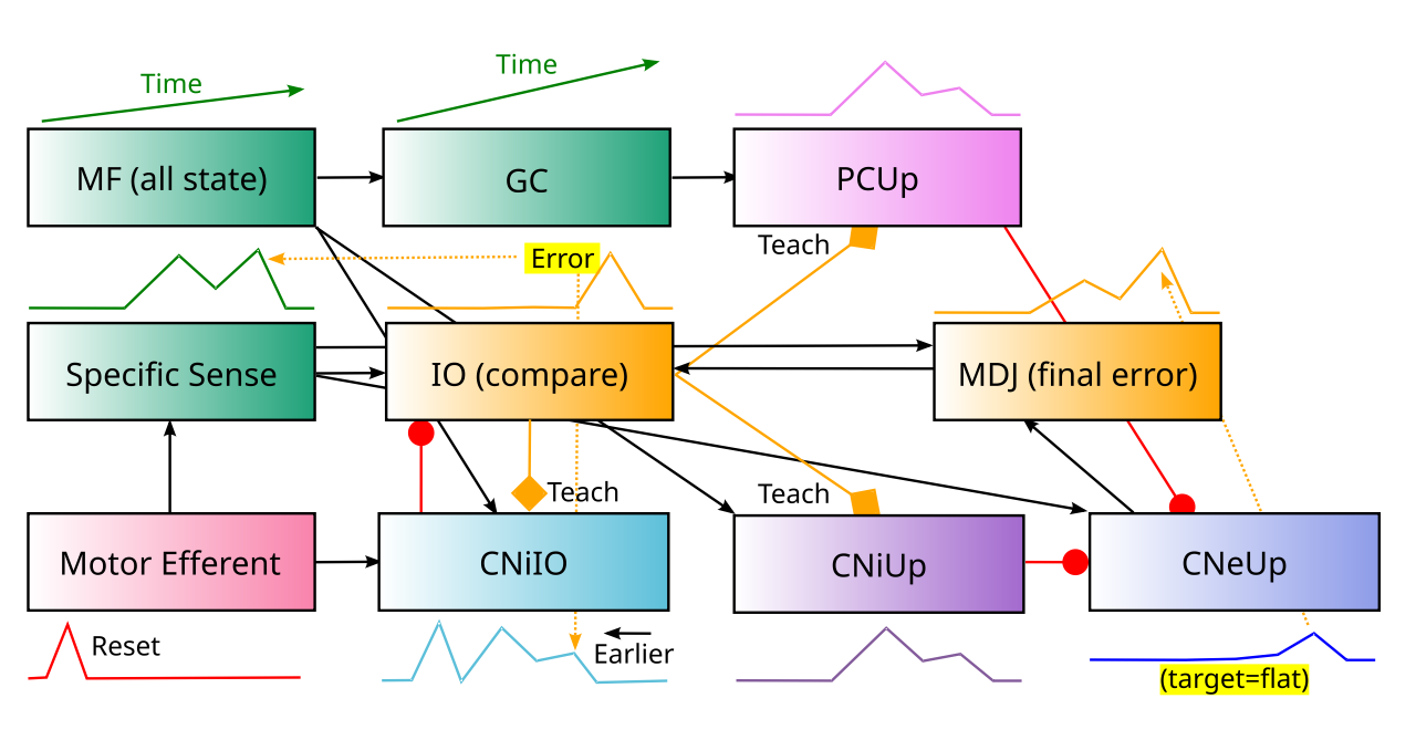
Figure 13: The mesodiencephalic junction (MDJ) nuclei receive excitation from CNeUp and project into the IO, so that IO error signals are also sensitive to this final output.Compare this figure with the adaptive filtering figure shown earlier.
Interestingly, the excitatory projections from upbound CNe neurons to the MDJ (mesodiencephalic junction) neurons, which in turn project to the IO neurons that project into corresponding CN microzones (Figure 13; De Zeeuw et al., 1998; Wang et al., 2022), provide a mechanism for direct error feedback learning based on the final CNeUp signal. Any residual perturbation in this signal can drive IO spiking, which should then drive further learning in the CNiUp neurons to better cancel the excitatory sensory inputs. This provides an important function for these otherwise rather mysterious MDJ areas.
Purkinje cells
The IO prediction error signal drives learning in the Purkinje cells (PC), which send a constant stream of GABA inhibitory simple spikes to the CNe output neurons. Thus, from the Nuclear perspective, these PC neurons have the same function as the CNi neurons, and the upbound vs. downbound learning dynamics in the PC neurons are consistent with the within-CN learning mechanisms described above.
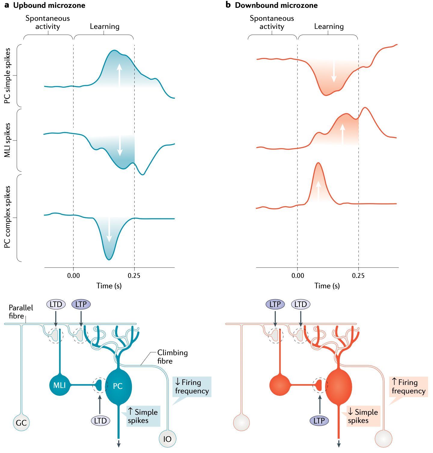
Figure 14: Plasticity in upbound vs downbound microzones, according to the framework of De Zeeuw (2021). In upbound microzones (which stain positive for zebrin II, also known as ALDOC), complex spikes driven by IO climbing fiber inputs drive learning that increases the rate of simple spike output (normal PC spiking activity). This is consistent with adaptive filtering, which needs to cancel out the predicted sensory signals, with increased PC inhibition. In downbound microzones, these complex spikes drive decreases in PC simple spiking, consistent with forward models that use anticipatory predictive signals to drive motor actions. The molecular layer interneurons (MLI; previously called stellate or basket cells) contribute to these patterns of plasticity. There are also differences in baseline firing rates associated with these different microzones (upbound have lower and downbound have higher).
Each IO neuron sends climbing fiber (CF) axonal output that goes up to the PCs (in addition to the CN pathway). Each IO neuron sends CF output to about 10 PC neurons, but each PC neuron only receives from one IO neuron, with about 300 synapses per PC neuron (Ito, 1984). The IO spikes drive powerful complex spike activity patterns in the PC neurons that trigger learning in these neurons (Ito & Kano, 1982; Coesmans et al., 2004; Gao et al., 2012; Silva et al., 2024), with the direction of learning determined by the microzone as shown in Figure 14.
In the adaptive filtering context (i.e., for upbound microzones), an IO-driven complex spike reflecting unpredicted sensory excitation should drive LTP (long-term potentiation) in the PCUp neurons, which would then increase the amount of inhibition they send to the CNeUp neurons, thereby balancing out the additional excitatory drive from the sensory input and canceling the sensory signal.
For the forward model case in the downbound microzones, the IO prediction error signal drives the opposite sign of synaptic plasticity in the PCDn neurons, resulting in a net disinhibitory effect on CNeDn neurons, which can then drive anticipatory motor actions. This opposite sign of PC plasticity corresponds to LTD occuring when there is an IO-driven complex spike, which is the pattern of plasticity predicted by the Albus (1971) and Ito (1972) motor learning theories.
This form of PC LTD has been extensively demonstrated and analyzed (Ito, 2001; Coesmans et al., 2004; Gao et al., 2012; Yang & Lisberger, 2014; Silva et al., 2024). It is notable that this downbound direction, needed for forward model learning, is the the most widely studied direction of plasticity, presumably because the motor actions that it supports are much easier to study than the adaptive filtering supported by upbound learning.
The fact that the cerebellar cortex has dedicated microzones for each direction of plasticity (De Zeeuw, 2021) is consistent with the differential connectivity demands for adaptive filtering vs. forward model functionality. Furthermore, the fact that one direction of plasticity is dominant in each microzone reflects the somewhat unusual nature of the IO climbing fiber training signal, which is naturally asymmetric, firing complex spikes only when the sensory input exceeds the CNiIO inhibitory prediction. Thus, the system appears to be organized to quickly respond to positive prediction errors (occurrences of unexpected sensory outcomes).
Thus, there remains a question as to what happens when expected predictions no longer occur, or the sensory prediction intensity exceeds the actual level. Empirically, there is evidence at multiple levels that PC and CN plasticity is bidirectional (Brooks et al., 2015; Brooks & Cullen, 2013), although it is generally slower in the “negative” direction (i.e., LTP for downbound, and LTD for upbound).
Indeed, the complementary direction is difficult to distinguish from a slow decay mechanism that is also necessary to accommodate the slower acquisition process taking place within the cerebellar nuclei. This is consistent with a systems-consolidation of learning from the rapid acquisition by the cerebellar cortex to the slower consolidation in the cerebellar nuclei.
Mossy fiber inputs
The mossy fiber (MF) inputs provide the “source” information for driving the predictive firing in the cerebellum. As such, they must provide information that is reliably predictive of the subsequent sensory and motor outcomes of actions. The most obvious source is the actual motor commands that are driving the behavior in the first place. These descending motor commands arise primarily from three major brain structures: the motor frontal cortex, the superior colliculus (SC), and the basal ganglia (BG), each of which project extensively into the pontine nuclei (PN) that provide the source of many of the mossy fibers. While the motor cortical projections are primarily via the PN, the SC and BG each have multiple routes for driving MF inputs.
The SC projects directly to the PN (Benavidez et al., 2021), and also to most of the other midbrain motor structures that also project into the CN, e.g., as discussed above in the context of the MDJ. As the primary source of evolutionarily shaped sensory-motor behavior, the SC inputs provide a critical early predictive signal about the motor commands driving behavior.
The projections from the basal ganglia originate from the subthalamic nucleus (STN) (Bostan et al., 2010) and the parafascicular nucleus of the thalamus (PF) (Marini et al., 1999), which provides a major representation of the BG output. These two areas represent the main excitatory areas that can communicate a positive representation of BG activity, with the STN having very early activity at the very start of the BG processing cycle (driven by neocortical and SC inputs; Coizet et al., 2009). By contrast, the PF reflects the output of the BG, which is also communicated directly via inhibitory projections from the SNr and GPi onto a wide range of subcortical targets, including the SC and midbrain motor areas.
Granule cells
The cerebellar cortex also contains a very large number of tiny granule cells (GC), that receive MF inputs and send excitation to the PCs, via distinctive parallel fiber axons, with each PC receiving 150,000 or more parallel fiber inputs. The large number of these GCs result in the cerebellum having about 70% of the total number of neurons in the human brain. These GCs, along with inhibitory Golgi cells and other specialized cell types in some areas, are thought to provide a very high-dimensional time-varying representation that is particularly useful for rapidly learning novel temporal firing patterns in the PC neurons (Marr, 1969; Buonomano & Mauk, 1994; Mauk & Buonomano, 2004).
Summary
In summary, the Nuclear model shows how the elaborate and specialized nature of the cerebellar circuitry seems ideal for performing the adaptive filter and forward model functionality, consistent with major longstanding hypotheses (Fujita, 1982; Miall & Wolpert, 1996; Bastian, 2006; Dean et al., 2010; Tanaka et al., 2020; Cullen, 2023).
The presence of two distinct pathways for canceling the sensory signal, a direct pathway within the CN and an indirect pathway through the cerebellar cortex, raises the question as to why two separate pathways would be useful, and how they might perform complementary functions? Taking inspiration from the seminal ideas of David Marr for both the hippocampus (Marr, 1971) and the cerebellum (Marr, 1969), one hypothesis is that the use of a very high-dimensional representational space in the cerebellar cortex (via the GCs) allows the system to rapidly learn new spatiotemporal firing patterns without suffering from extensive interference with prior learning.
Furthermore, the CN system can slowly learn the most reliable and stable forms of sensory predictions, while the cerebellar cortex can rapidly learn more context-dependent, idiosyncratic predictions, which may then decay away over time (Herzfeld et al., 2020; Kimpo et al., 2014; Kassardjian et al., 2005). In short, the CN slowly learns the rules, while the cerebellar cortex can quickly learn the exceptions, just as in the complementary learning systems framework for the hippocampus vs neocortex (McClelland et al., 1995; O’Reilly & Rudy, 2001; Norman & O’Reilly, 2003).
There is evidence that the CN pathway does not have as accurate of timing relative to the cortical pathway (Garcia et al., 1999), which is consistent with the high-dimensional representation formed in the cerebellar cortex. Because each PC integrates input from roughly 150,000 granule cell inputs, it can form novel, arbitrary associations across many more diverse stimulus inputs than the more limited dendritic arbors of CNiIO neurons. Thus, the CN likely learns coarser timing while the cerebellar cortex learns fine-grained timing. Where this fine-grained timing remains persistently relevant, continued error signals could sustain the cortical pathway contributions, with a stabilization process over time making these connections permanent over the longer term.
A number of studies are consistent with rapid one-trial learning in the cerebellar cortex (Medina & Lisberger, 2008; Kimpo et al., 2014; Yang & Lisberger, 2014; Herzfeld et al., 2020). These studies also demonstrate the rapid decay of the strongest synaptic changes that occur on a single trial, but are also consistent with considerable evidence of long-lasting changes in the cerebellar cortex synapses as well (Silva et al., 2024; Ito, 2001; Wang et al., 2014).
There is a significant mismatch between the stimulation paradigms used in most synaptic plasticity studies conducted in slices (Ito, 2001; Ito, 2013) and those demonstrating rapid but transient changes in awake behaving animals (Medina & Lisberger, 2008; Kimpo et al., 2014), which could account for the lack of focus on these dynamics in the broader cerebellar synaptic plasticity literature.
Learning rules
There is biological evidence for synaptic plasticity in just about every synapse in the cerebellar system (Hansel et al., 2001; Carey, 2011; Gao et al., 2012). The most important synapses for this plasticity based on the logic of Figure 11 and Figure 12 are the MF mossy fiber inputs to the CNiIO, CNiUp, and PC neurons, which each need to learn a phasic, anticipatory pattern of activity.
There is a general consensus in the field that initial faster learning in the PC neurons is necessary for the systems consolidation of slower learning in the CN (Raymond & Medina, 2018; Herzfeld et al., 2020). However, each of the relevant studies supporting this conclusion has issues that leave this point in doubt, suggesting that learning in the CN can proceed independently from the PC inputs, but that these PC inputs may also facilitate learning when present.
Perhaps the strongest-seeming data comes from Garcia et al. (1999), who lesioned the cerebral cortex post-learning in an conditioning paradigm, and found that this lesion prevented subsequent acquisition to a new CS. However, this is could actually have been due to a blocking effect from the prior CS-US conditioning, as was found by Kim et al. (1998).
Another study by Zeeuw et al. (1998) used a genetic knockout to disable a critical protein kinase involved in synaptic plasticity, selectively in the PC neurons. Although they found that this blocked adaptation of the vestibulo-ocular reflex (VOR) in both the PCs and the CN, they only used a short amount of training (1 hr) which was shown in another study to only affect learning in the cerebral cortex (Kassardjian et al., 2005).
Consistent with the fast vs. slow dissociation, Kassardjian et al. (2005) only found cortically independent learning in the CN after 3 days of training. Furthermore, Zeeuw et al. (1998) noted that the standard VOR, and considerable motor behavior, was intact in their knockout mice, providing strong evidence that plasticity in the PC is not important for the slow learning over developmental time in the CN.
Another study by Shutoh et al. (2006) used a range of manipulations and found that the inactivation of the IO significantly impaired longer-term VOR adaptation, consistent with the idea that IO is critical for learning in the CN neurons. Inactivations and pharmacological manipulations of the cerebellar cortex consistently had less of an effect than those of the IO, but still had significant effects, consistent with an additional role of PC to CN projections in learning within the CN. This is potentially inconsistent with the Zeeuw et al. (1998) study, and further research is needed to resolve these discrepancies (see also Ito, 2013). Nevertheless, it seems reasonable to assume that CN neurons can learn independently in conjunction with IO input, and that the PC inputs provide additional learning signals based on their own rapid learning.
Predictive delta rule
At a computational level, the simple delta-rule function is optimal from a mathematical perspective for learning to predict a sensory input signal s in the activity of a receiving neuron y (as in the CNiIO neurons):
\[ \Delta w = (s - y) x \]
where x is the sending activity (from mossy fiber inputs).
This equation requires bidirectional learning based on the mismatch between the sensory value and prediction: if the prediction is too low, then the synaptic weights need to be increased (LTP), and decreased (LTD) in the opposite case. There is direct evidence for bidirectional synaptic plasticity in CN neurons generally (Aizenman et al., 2000; Pugh & Raman, 2008; Pugh & Raman, 2006; Zhang & Linden, 2006; McElvain et al., 2010).
However, the predictive nature of the cerebellum introduces a further challenge, which becomes apparent when we add time to the learning rule, with \(\epsilon\) representing the number of ms in advance that the prediction \(y\) anticipates (e.g., 80 ms):
\[ \Delta w_t = (s_t - y_{t-\epsilon}) x_{t-\epsilon} \]
(equivalently, you can add \(\epsilon\) to all of the time indexes in Eq 2).
Thus, the error term (\(s_t - y_{t-\epsilon}\)) needs to be sensitive to the predictive temporal offset, so that the prediction activity at time \(t\) is about the sensory input that will arrive e.g., 80 ms later (i.e., anticipating the later input). The natural trace-like integration process in the inputs can handle the \(x_{t-\epsilon}\) factor for the sending inputs (Raymond & Lisberger, 1998; Wang et al., 2000), but computing an error term that is offset across time actually requires some kind of delayed comparator process.
As discussed above, the IO neurons have specialized neural machinery that can perform exactly this kind of temporally offset comparison function, where earlier inhibitory predictive inputs from the CNiIO neurons are needed to block the impact of excitatory sensory inputs that arrive roughly 50-100 ms later. This suggests that learning in the CNiIO and other CN neurons should be dependent on error signals coming from the IO neurons, just like the PC neurons are, which is consistent with the presence of collateral projections from the IO into the CN (Want & Voogd, 1987; De Zeeuw et al., 1997). Thus, we need to understand the computational properties of this IO signal.
Properties of the IO error signal
The widely accepted computational-level idea that the IO provides an error signal that drives learning in the cerebellum has been difficult to reconcile with the actual properties of neural firing in IO neurons (Najafi & Medina, 2013; Zang & De Schutter, 2019). They fire regularly at low rates, roughly 1 hz, which is a seemingly impractical baseline level: it is too low to provide much of a dynamic range for reductions in firing rate to be meaningful, and yet it is high enough to reduce the signal-to-noise ratio of IO activity for representing errors (Llinás et al., 1997). If IO activity means an error occurred, then there is a false alarm every second! But the absence of this once-a-second IO firing could only provide an unhelpfully wide timing window to interpret this absence as a meaningful signal.
As a potential mitigation of the issues with tonic background spikes, there is evidence that the firing pattern associated with an actual IO error signal is distinct from the baseline 1 hz firing. One set of studies showed a relationship between oscillatory phase in IO neurons and the number of small bursts triggered by a single initial IO action potential (Mathy et al., 2009), which could potentially discriminate signal from the background. More recently, a stimulus input was shown to drive a pair of IO spikes within around 100 ms of each other, which results in more robust synaptic plasticity in PC neurons compared to a solitary spike typically generated by the background rate (Titley et al., 2019).
However, these paired IO spikes were not observed in a recent in vivo calcium imaging study, which instead showed more synchronous population activity across multiple IO neurons for stimulus-driven IO activity relative to the 1 hz baseline (Guo & Uusisaari, 2025). In any case, given the strong in vivo neural recording evidence that PC activity is bidirectionally modulated (Coesmans et al., 2004; Gao et al., 2012; Kimpo et al., 2014; Yang & Lisberger, 2017), it seems likely that there is some way in which the true error signal has distinct effects from the background spikes.
Two key considerations can help understand why the IO error signal has the properties it does. First, the precise timing of the learning signal is of paramount importance, and timing precision is directly conflicting with gradedness for biochemical processes that unfold over time. If there is a single spike, then learning processes can proceed immediately at the point at which that spike arrives, reflecting the state at that time. However, to communicate graded quantities, multiple spikes need to be sent over time, meaning that by the time the graded quantity is established, a potentially variable amount of time has passed, and the state of the rest of the system will have thus moved on.
This single precisely timed spike case also then requires that the error signal be asymmetric, given that this one spike has only a single bit of information, which can either signal when the target value has not been sufficiently predicted, or when the prediction exceeds the target value. Of these two, signaling the onset of an unexpected sensory input is definitely more important.
Second, learning in both the CN and the PC neurons does not actually require bidirectional or graded error signals, for different reasons in each case. For CN neurons, they learn very slowly, so single small increments on each learning trial are sufficient, and the additional value of a graded signal on each trial is minimal. For PC neurons, they learn quickly, and, critically, do not actually alter the CNiIO prediction that controls IO firing. Therefore, they need to saturate automatically, and the graded value of the error is not actually meaningful.
For both neuron types, a bidirectional signal is not necessary because they can each use decay (slowly for CN, more rapidly for PC) instead of bidirectional error signals. Decay in the CNiIo prediction neurons will result in occasional error spikes that bump the synaptic strengths back up as the lower limit is tested, while decay in the PC neurons allows the system to more quickly fall back on the slower CN learning, which is important to do in any case. It is likely that the background 1 hz spiking provides an opportunity to drive a slow decay process.
Thus, in light of these considerations, the IO error signal starts to make more sense. Furthermore, even the 1 hz background firing rate makes sense in terms of the full time window over which the temporally offset comparison process operates. Specifically, because the earlier prediction signal needs to be compared against a sensory signal arriving around 50-100 ms later, and the window for relevant sensory signals around a given motor action could spread out for 500 ms or more, the entire temporal envelope of an ongoing comparison process (Figure 9) could stretch to upwards of a second in total. Thus, perhaps the 1 hz baseline firing rate reflects this action envelope for the comparison process, which is reset every second, and is synchronized by the initial descending action signal and the gap junction connections within the IO glomeruli.
IO-driven learning rule
The final step to define the cerebellar learning rules is to translate Eq 2 into equations that operate when there is an IO error signal, and when there is a baseline spike that drives synaptic decay:
\[ \Delta w = \lambda_e x_{t-\epsilon} \; \; \rm{error} \]
\[ \Delta w = -\lambda_d x_{t-\epsilon} \; \; \rm{decay} \]
where \(\lambda_e\) is the learning rate for error trials, and \(\lambda_b\) is the decay learning rate. Note that the decay learning operates on whatever inputs are active at the time of the baseline 1 hz spike that triggers decay learning, instead of generically decaying everything. For the PCDn learning, the signs are reversed.
In support of the asymmetrical directionality of this learning rule, there are well-documented cases across multiple paradigms that show strong such asymmetries. For example, in a very thorough study of the VOR (vestibulo-ocular reflex) paradigm, only increases in VOR gain exhibited rapid trial-by-trial adjustments, that were consistent with LTD of the PC climbing fiber synapses (Kimpo et al., 2014). Nevertheless long-term changes in behavior and PC firing were appropriately bidirectional, suggesting that the asymmetry was restricted to rapid, transient changes. There may be general functional benefits to this asymmetry that can be investigated with computational models.
A directional saccade task also showed the same kind of asymmetry in rapid learning, which was also dissociated from the bidirectional plasticity in longer-term learning (Yang & Lisberger, 2014). The widely studied eyeblink conditioning task has shown more ambiguous results on the directionality of learning (McCormick & Thompson, 1984; Berthier & Moore, 1986; Jirenhed et al., 2007; Green & Steinmetz, 2005), perhaps consistent with a mix of both forward model and adaptive filter modes of learning.
Also, there are a number of additional factors that have been identified in driving plasticity in the overall PC simple spike responses, including changes in the nature of the mossy-fiber inputs, and plasticity in the molecular layer interneurons that inhibit PC neurons (Bonnan et al., 2023; Zhang et al., 2023). Some of these factors likely modulate PC learning so it only occurs in response to meaningful IO spikes and not to the regular 1 Hz background spiking (Zang & De Schutter, 2019; Guo & Uusisaari, 2025).
Standard models
There have been a large number of different theoretical accounts of cerebellar function over the years (see Nguyen & Person, 2025; Shadmehr, 2020; Raymond & Medina, 2018 for recent reviews). Most of these models have focused on the circuits in the cerebellar cortex, with relatively little attention paid to those in the CN (Kebschull et al., 2024; Herzfeld et al., 2020). In this section, we review some key points of divergence among the various theories, in relation to the Nuclear framework presented above.
At a broad level, the adaptive filtering and forward model functionality supported by the Nuclear model is consistent with longstanding and widely discussed principles of cerebellar function (Fujita, 1982; Miall & Wolpert, 1996; Bastian, 2006; Dean et al., 2010; Tanaka et al., 2020; Cullen, 2023). Nevertheless, the Nuclear model provides a biologically specific, mechanistic account for the origin and nature of the predictive error signal in the IO, and for the roles of the different neurons within the cerebellar nuclei, that have not been fully integrated in prior accounts.
De Zeeuw (2021) provides examples of upbound and downbound learning in various well-studied domains, and argues that the number of inhibitory synapses involved determines the directionality (odd number = upbound, even number = downbound). However, he does not directly connect this distinction to the adaptive filtering vs. forward model frameworks. In addition, the detailed CN circuitry that is needed to allow consolidation of initial PC-based upbound and downbound learning was not specifically addressed in the De Zeeuw (2021) proposal. This machinery, along with the unique role of the MDJ (mesodiencephalic junction) in the upbound microzones, goes well beyond the difference in output polarity that is emphasized by De Zeeuw (2021).
Interestingly, recent research shows that the strong spatial organization of the upbound vs. downbound microzones might be specific to rodents, with primates showing a more intermixed structure (Hao et al., 2024), which could explain various differences across these species (e.g., compare Kimpo et al., 2014 in monkeys vs. Voges et al., 2017 in mice).
Perhaps the greatest source of remaining uncertainty and overall confusion in the literature concerns the role of the cerebellum in more flexible, open-ended motor control. Many have assumed that the cerebellum can manage core aspects of this functionality all on its own, which then requires considerably more complex and difficult computational problems to be solved (e.g., Kawato, 1999, Llinás, 2009). However, many lines of evidence strongly suggest that such abilities require the coordinated interactions among multiple brain areas including the basal ganglia and the prefrontal cortex, as reviewed in the motor page. Thus, by putting the cerebellum into a broader context of different neural systems, each specialized for one well-defined type of neural computation and learning, we can perhaps arrive at a more satisfying overall understanding.
Nature of the IO error signal
It is interesting that although most discussions of cerebellar function depict the IO as performing an error computation, and a number of papers discuss both the prediction error coding and these detailed biological properties (e.g., Loyola et al., 2023; De Zeeuw et al., 1998), none have articulated the direct and explicit connection between the unique biological properties of the IO neurons and this error computation.
The summary of the IO error signals and learning across the many eyeblink conditioning studies provided by Ohmae & Medina (2015) has been highlighted as demonstrating a novel form of error computation in the IO (Hull, 2020; Nguyen & Person, 2025). However, these properties are all consistent with the delta rule learning algorithm hypothesized for the CNiIO prediction learning pathway (Eq 1). This equation is otherwise known as the Rescorla-Wagner learning rule (Rescorla & Wagner, 1972), originally used to explain a wide range of conditioning phenomena associated with phasic dopamine firing (see PVLV for further discussion). The only aspect of this learning that is potentially challenging is the CS-based responding, but this is consistent with the anticipatory bias of cerebellar predictive learning.
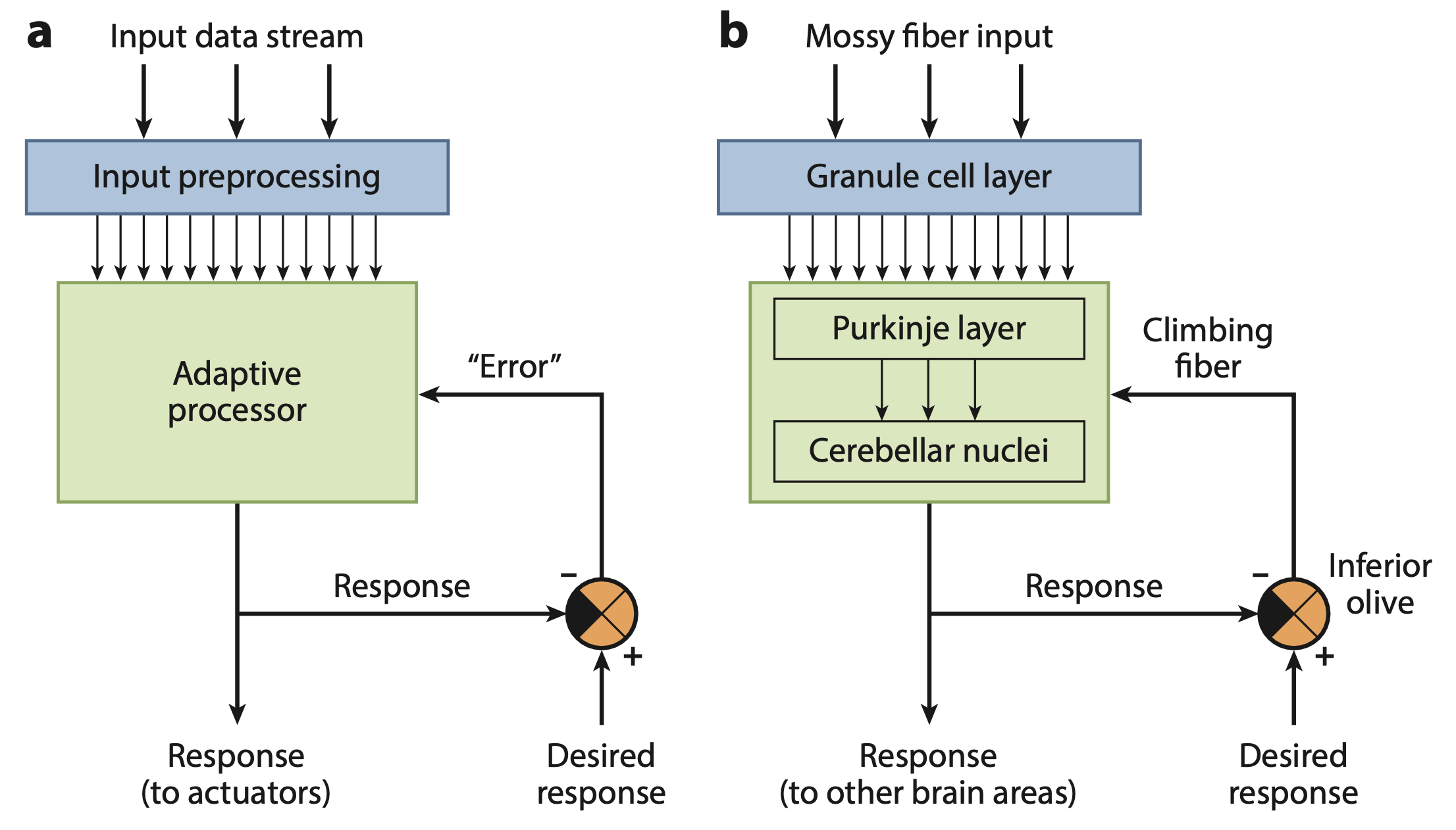
Figure 15: Diagram of cerebellar function from Raymond & Medina (2018), showing the critical IO error signal computed as a difference between the actual response and a “desired response”. The origin and nature of this “desired response” is unclear, representing a critical weakness in most standard accounts of cerebellar function.
In contrast to the well-defined, biologically explicit mechanism in the Nuclear model for computing a sensory prediction error, most other treatments of this essential aspect of cerebellar function are notably vague, as illustrated in Figure 15 from Raymond & Medina (2018). How and where does the brain compute something as omniscient as a “desired response”? And if such a thing is available, why not just use it directly, instead of making undesired responses in the first place?
Indeed, a necessary step for making the error signal directly computable is to define it in terms of sensory outcomes, because these are the concrete outcome signals that project into the cerebellum. However, because the cerebellum is involved in motor control, people naturally want to think in terms of motor errors, not sensory errors. This is where the problems arise: it is really unclear how to define a “motor error”, as discussed next.
Motor space is messy, sensory is clean
The fundamental problem with motor control, and thinking in terms of representations in motor control space, is that it is a very messy place. Specifically, there is a massive one-to-many relationship between the desired outcome of a motor action and the actual muscle movements necessary to accomplish that outcome, depending on a huge number of variables that are generally different for each such action. Consider reaching for a cup on the table. The precise motor actions needed to successfully accomplish this simple action depend on the initial position of each joint in your arm, the postural configuration of the rest of your body (e.g., standing or sitting, and exactly how in relation to factors such as balance), the physical properties of the cup, the fatigue state of your muscles, etc. Each of these factors interact, creating a massive combinatorial explosion problem.
For this reason, it is much simpler to drive motor control in sensory space (Powers, 1973), where both the targets of motor actions and their outcomes are all encoded in a much lower-dimensional, more systematic representational space. Thus, when you reach for the cup, the visual, somatosensory, and proprioceptive signals tell you whether the action was successful, and in what direction it was off, etc.
Furthermore, the adaptive filtering process can systematically clean up the sensory space, so that all of the factors mentioned above (starting joint angles, posture, balance, etc) have already been subtracted from the relevant sensory signals. Therefore, the motor planning based on this filtered sensory space is much more systematic and lower-dimensional.
This sensory-based framework contrasts with the attempt to invert a forward model to obtain an inverse model (Jordan & Rumelhart, 1992; Kawato, 1999), which in principle tells you what motor commands are needed to obtain a given sensory outcome (recall that the forward model goes the other way, predicting the sensory outcome of a motor command). For all the reasons discussed above, it is quite difficult to create an inverse model in complex, real-world cases, because there are often many possible action sequences that could result in the same or similar outcomes (Jordan & Rumelhart, 1992; Gibson et al., 1997).
Further insight into problems with motor-based frameworks comes from a consideration of the predictive error signals computed by the IO, which have been proposed to be based on efferent copy inputs (Oscasson, 1980; Kawato & Gomi, 1992). Simpson et al. (1996) note that the inputs tend to be dominated by somatosensory and proprioceptive signals, and not efferent copy inputs, and that there is a timing issue for motor error computation, where the temporal separation between the initial motor command efferent copy and the subsequent motor outcome can be relatively long (more than 100 ms, as shown in Figure 9). Furthermore, there is an anatomical mismatch between descending and ascending motor pathways as they map into the cerebellum (De Zeeuw et al., 1998).
Pathways not included in model
The following pathways have been documented but are not included in the present model (and generally these are not widely discussed).
- • Uusisaari & Knöpfel (2010) demonstrated a projection from glycinergic (inhibitory), not tonically active neurons in the CN that project into the cerebellar cortex. These were relatively sparse and late-maturing.
Summary
In summary, sensory predictive learning (forward models) and adaptive filtering provide a fully computationally well-defined framework that is consistent with known cerebellar properties, as captured in the Nuclear model. If it is possible to implement functional motor control using just this adaptive filtering functionality to provide high-quality sensory signals upon which motor actions can be based, then many otherwise challenging difficulties disappear. The broader framework provided in the motor page, and implemented computational models that include the function of the basal ganglia and prefrontal cortex, demonstrate that this is indeed possible.
Furthermore, the Rubicon goal-driven control framework provides a critical outer-loop infrastructure for motor action selection to accomplish desired goal outcomes. This system uses a sensory-based goal selection mechanism, informed by multiple levels of predictive learning systems and shaped by constraint satisfaction processing across learned distributed representations, to select a desired outcome and an overall plan of action. This goal-selection process can be informed by cerebellar predictive learning mechanisms, but does not require inverting forward models to obtain the inverse model.
Vestibulo-ocular reflex
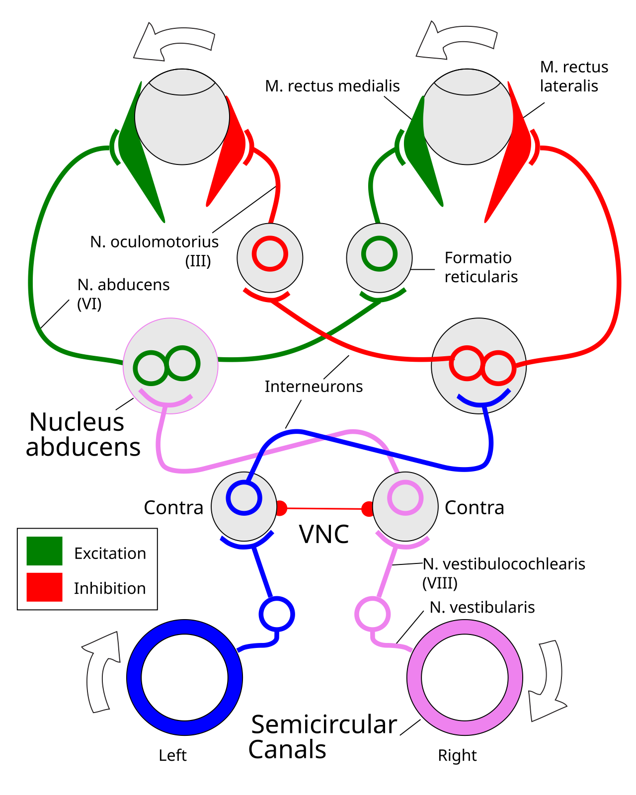
Figure 16: Anatomy of the VOR reflex, which is driven by vestibular signals from the semicircular canals, which project into the vestibular nuclear complex (VNC), and from there into optical motor areas. All primary inputs are ipsilateral (e.g., the right ear projects to the right hemisphere), and the VOR involves the subset of VNC neurons that project contralaterally to drive the nucleus abducens optical motor interneurons that cause eye movents in the compensatory, opposite direction. There are also ipsilateral pathways, which are inhibited by the contralateral ones, as well as cross-hemisphere inhibitory competition. This is a classic opponent process organization. The VNC neurons in this circuit are the functional equivalent of the cerebellar nucleus neurons in other areas, and thus direct plasticity in therse neurons is the ultimate consolidated result of learning. The cerebellar-cortex modulation of the gain on this reflex arises from changes in the tonic neural activity onto the VNC neurons from Purkinje neurons (e.g., disinhibiting increases gain), and in synaptic inputs to these VNC neurons. Adapted from wikimedia.org.
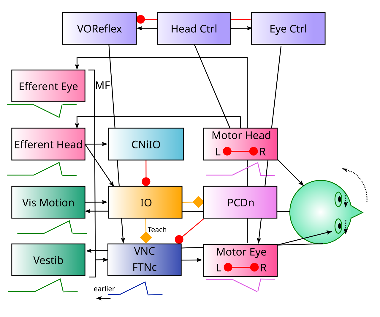
Figure 17: Central elements of VOR gain modulation in the cerebellum. The violet colored boxes at the top reflect brainstem control areas where descending cortical and basal ganglia motor inputs drive coordinated motor behavior. The Head Ctrl area drives neck rotation, but also by default provides inputs into the VOReflex motor reflex area that modulates the VOR compensatory eye movement circuitry (as shown in prior figure). The Eye Ctrl area supports direct saccadic control over eye position, which automatically suppresses the VOR, by inhibiting the VOReflex. The CNiIO (predictive inhibition) and visual motion (specific sensory input) drive error spiking in the associated IO neurons, which then drive learning in the FTNc (flocculous target neurons, contralateral) in the VNC (vestibular nuclear complex), which are the functional equivalent of the cerebellar nucleus (CNeDn) neurons. Floccular Purkinje neurons (PCDn) also learn (LTD, downbound) from the IO and thus disinhibit these FTNc neurons.
The vestibulo-ocular reflex (VOR) is a thoroughly studied aspect of cerebellar function (see Cullen, 2023; Kheradmand & Zee, 2011 for reviews, and Zeeuw et al., 1998; Kimpo et al., 2014 for important results). It provides a well-understood example of the forward model, downbound aspect of cerebellar function, where the onset of a head movement triggers anticipatory eye movements to compensate for that head movement, by moving in the opposite direction. The net result should be a relatively stable image on the retina, and the global visual motion signal (i.e., retinal slip) drives the IO error signal that then drives cerebellar learning.
The VOR is directly mediated by brainstem circuits driven by vestibular sensory inputs (Figure 16). In this case of the evolutionarily most ancient vestibular system aspect of cerebellar function, the functional equivalent of the cerebellar nucleus neurons are located directly within the secondary vestibular nuclear complex (VNC), as shown in the figure. These neurons directly participate in the VOR reflex, whereas in other cases the cerebellar nucleus neurons would typically be modulatory onto other brainstem circuits, e.g., in the MDJ and other areas.
The reflexive circuit shown in Figure 16 is typical of many similar such circuits in different parts of the brainstem, where sensory signals directly drive associated motor behavior, via secondary neurons that can be modulated to enable or disable the reflex.
Although it is described as a reflex, the key cerebellar contribution to the VOR is to anticipate (through downbound forward model pathways) the sensory effects of head motion, so that the compensatory eye movements are not always lagging behind, and instead can be anticipatory and more precisely negate the effects of head motion.
Logically, this forward model would be one that is based on the actual full-field visual motion signals from the retina that would otherwise be produced by the head motion, because those will have the relevant temporal envelope for the actual motion signals that are being canceled. However, there is a catch-22 associated with those signals: they will be zeroed out by the compensatory eye movements. Therefore, it is important that the specific driving sensory input that is being anticipated comes from the vestibular senses, which will always be activated by head motion even with the active VOR eye movements.
In experimental studies, the contribution of the cerebellum is summarized by its contributions to the VOR gain factor: the ratio of head motion to compensatory eye motion in the opposite direction. For the normal, baseline case, this gain factor is 1. But experimental manipulations including changing the magnification in the eyes (via glasses) or stimulating the vestibular nerves causes the gain to change, in a cerebellum-dependent manner.
The VOR gain is a multiplicative factor, and yet neurons generally can not directly drive multiplicative effects on other neurons. Instead, the way this is accomplished in these circuits is through an opponent organization, so that the modulatory neuron excites both a pull towards and a push away from the target action. As the level of excitatory drive is modulated, the overall range of firing of these opponent neurons will vary as well, and it is this overall range that corresponds to the effective gain factor.
TODO: key question of how zero visual motion as error signal translates into learning – this zeroing out is probably true for many other cases..
Some of the relevant phenomena that a biologically based model should account for includes:
• The asymmetry in VOR gain increases vs. decreases, in terms of rate of learning and duration of retention (Boyden et al., 2004; Kuki et al., 2004; Kimpo et al., 2014). We can understand these differences in terms of the rapid contribution of downbound cerebellar cortex LTD learning for gain increases, which decays relatively rapidly, versus slower, longer-lasting changes in the effective cerebellar nucleus neurons (Lisberger et al., 1994).
• Blazquez et al., 2003 document a number of different changes in the responses of Purkinje cells to various input parameters, which together can account for the VOR gain changes.
• Detailed patterns of changes in secondary vestibular neurons that mediate the VOR, known as floccular target neurons (FTNs), including those with ipsilateral vs. contralateral firing properties, as documented by Lisberger et al. (1994) (e.g., Figure 18).
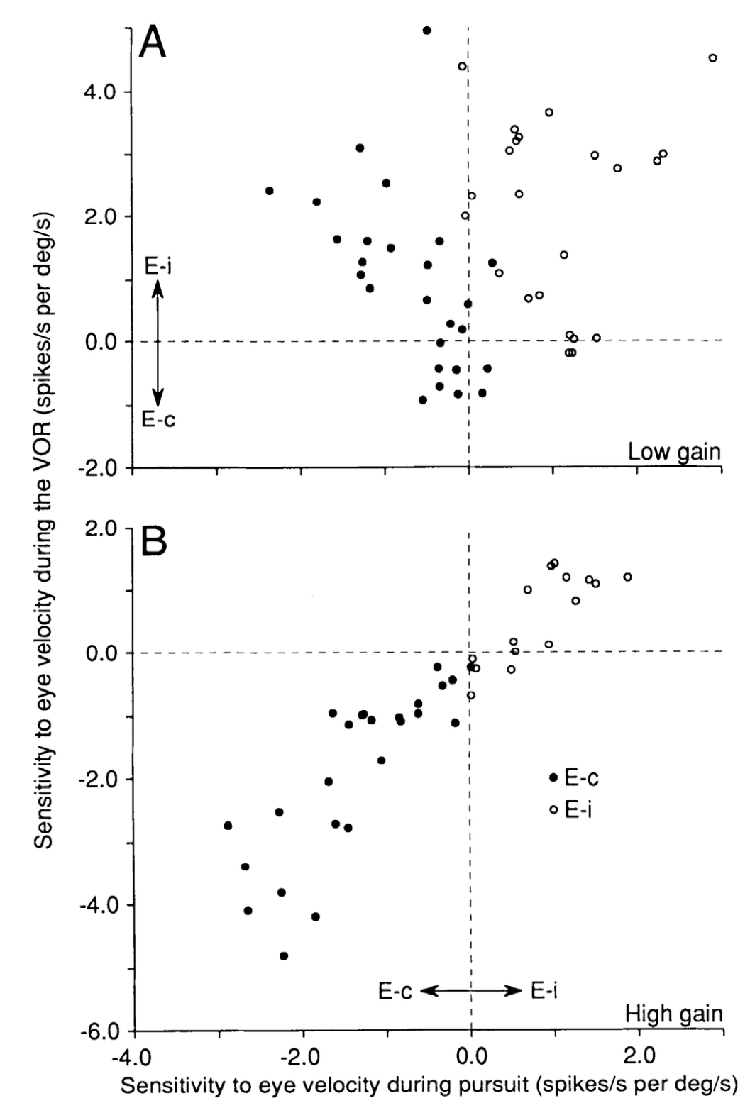
Figure 18: Results from Lisberger et al. (1994) on the modulation of FTN (floccular target neurons) in the vestibular nuclear complex (VNC). The E_i neurons have ipsilateral coding, while the E_c are the primary contralateral coding neurons that are thought to be the primary target of cerebellar modulation, with the effects on the ipsilateral neurons arising from inhibitory competition due to the opponent organization of this system.
Most of the existing models are relatively abstract (e.g., Holland et al., 2020; Inagaki & Hirata, 2017, Clopath et al., 2014; Lisberger, 2009; Kawato & Gomi, 1992; Robinson, 1981) and none appear to provide a detailed mechanism by which the IO comes to compute the critical training signal that drives learning in the Purkinje and vestibular nucleus cells. However, aside from that critical distinction, the Nuclear model that you can explore in VOR simulation is overall consistent with the principles established by these existing models.