
Thalamus
The thalamus plays essential roles in multiple different brain circuits, and is thus discussed mostly in the context of those other circuits. This page provides a big-picture organizational and functional view of the thalamus, with pointers to those other pages. Understanding the functions of the thalamus provides a powerful unifying and bridging tool for understanding neural function more broadly, because once you understand how one domain works, the same concepts can be extended to other domains.
From an evolutionary, comparative perspective, the thalamus consistently provides interconnectivity between primary sensory areas and the pallium, which is the outer-most region of the brain, i.e., the telecephelon. In mammals, the neocortex is a major component of the pallium, while other vertibrates have smaller, simpler cortex-like structures in their pallium that play the same functional role as the neocortex (Butler, 2008). The following functions are specific to mammals:
• Relaying with attentional modulation the primary sensory and motor inputs to the neocortex, such as the LGN (lateral geniculate nucleus) for vision. There are extensive top-down projections from sensory cortical areas back to these primary sensory thalamic areas, supporting attentional modulation of these sensory inputs (Poltoratski et al., 2019; Briggs, 2020). Primary motor cortex receives both a direct primary motor input from the cerebellum and inputs from somatosensory thalamus, which play an analogous role on the motor side, teaching motor cortex how to “speak in the language of the spinal cord”.
• In posterior neocortex, the pulvinar nucleus of the thalamus supports predictive learning by alternatively representing a top-down prediction and a bottom-up outcome. Similar connectivity is present in a subset of neurons within the mediodorsal (MD) nucleus that interconnects with prefrontal cortex and lower motor cortex areas, suggesting a similar predictive learning role. Likewise, anterior thalamic areas connected to the brain systems processing space can support this form of learning.
• There are multiple different thalamic nuclei including the AM, MD, and VA / VM that receive disinhibitory output signals from the basal ganglia (BG) and project collectively to all areas of motor and prefrontal cortex (PFC), where they can drive rapid phasic changes in activity states (i.e., gating; see the PBWM model), such as those evident during the transition from motor planning to actual execution. In the core goal-driven areas of PFC, these gating signals drive the transition from goal selection to the goal-engaged state, as captured in the Rubicon model. There is also an important subset of predictive learning neurons in these frontal areas that integrate predictive learning with BG gating, known as the IT layer 5 neurons (Shepherd & Yamawaki, 2021).
• The intralaminar (IL) nuclei provide an extensive feedback pathway for the basal ganglia.
• The paraventricular thalamic nuclei interconnect with the goal-driven PFC, hippocampus and associated emotion processing subcortical areas, and provide overall modulation of goal-driving processing.
Thalamic circuitry
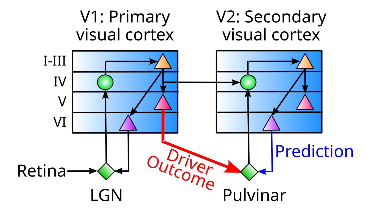
Figure 1: Connectivity between the neocortex and the pulvinar nucleus of the thalamus, in the case of primary and secondary visual areas. Visual input from the retina provides driver inputs to the lateral geniculate nucleus (LGN) of the thalamus, which then sends glutamatergic excitatory projections primarily into layer 4 of primary visual cortex (V1). The pulvinar thalamic nucleus receives strong driver inputs from deep layer 5b neurons in V1, which can have as few as one driver synapse per thalamic relay neuron (TRN). There are numerous and relatively weaker projections from layer 6 (VI) neurons in secondary visual cortex (V2). Diagram based on Sherman & Guillery (2006).
Figure 1, based on Sherman & Guillery, 2006, shows the thalamic connectivity for primary and secondary visual cortex in the mammalian brain. The main features of this connectivity are common across most thalamic areas.
There is only one predominant type of thalamic neuron in the different thalamic nuclei, the thalamic relay neuron (TRN). The thalamic reticular nucleus (TRN), which is an outer shell wrapping around the body of the thalamus, is the one exception, having GABAergic inhibitory neurons, which we discuss below. TRNs release excitatory glutamate neurotransmitter into the neocortex, and receive two qualitatively distinct types of inputs:
• Drivers are strong inputs, often relatively few in number (as few as a single input). They can have large extended glomeruli with multiple synaptic connections and even a “dedicated” inhibitory intgerneuron encapsulated in a single anatomical structure. These can originate from subcortical areas (e.g., retinal input to the LGN for vision) or cortical areas (e.g., the deep layer 5b IB intrinsic bursting neurons that are drivers in the predictive learning performed by the pulvinar). Some thalamic areas have no identified driver inputs, and are typically those that receive extensive inhibitory inputs from the basal ganglia output pathways.
The nature of the driver inputs is one of the most important clues to thalamic (and cortical) function. For example, cortical drivers from intrinsic bursting neurons provide the temporal dynamics needed to support predictive learning via the temporal derivative mechanism, whereas subcortical sensory drivers do not appear to have this property. However, subcortical drivers in the motor system naturally have the requisite delays, as discussed in the motor page.
• Non-drivers are the rest of the inputs, which are like most other synaptic connections in the brain. Sherman & Guillery, 2006 and many others label these as modulatory inputs, but that is perhaps too strong of a functional assumption, given their proposed function in the context of predictive learning. For example, there is ample evidence that these inputs can drive sustained firing of TRNs, as reviewed in O’Reilly et al., 2021.
The TRNs notably lack any local excitatory interconnectivity, and only have a form of pooled inhibition mediated by the TRN reticular neurons. Therefore, they are uniquely suited for providing a faithful representation of the inputs they receive, serving as a kind of projection screen that does not impose significant distortion of its own.
Although the relatively inert nature of the thalamic circuitry has led to its characterization as a relay system, the presence of two distinctive types of inputs suggests a more powerful kind of function could be be supported. The predictive learning framework for example shows how the distinctive temporal aspects of the driver inputs to the pulvinar supports a temporal derivative computation that effectively computes an error gradient between the driver and non-driver inputs, across time.
The nature of the connectivity from TRNs into the cortex provides another important insight into the function of the thalamus. There are two primary dimensions along which these projections vary, which were originally captured by the core vs. matrix categories of Jones (1998), but which are more accurately categorized as dimensions (Clascá et al., 2012; Sherman & Usrey, 2024):
• Laminar targets: the classical core type projects mainly to the middle layers of the cortex, primarily layer 4 (e.g., as shown in Figure 1), while the classical matrix type projects to layers 1 and 5, but avoids the middle layers. However, there are actually different proportions of these connections across different thalamic neurons, rather than sharply defined categories.
Functionally, projections into the middle layers provides direct input information into the cortex, as in the prototypical sensory input into primary sensory areas. Projections into layers 1 and 5 can drive two main functions: regulation of overall cortical activity levels via the VIP inhibitory interneurons in layer 1 (this was the original interpretation of the matrix type), and maintenance of sustained activity (primarily in prefrontal cortex) via bidirectional excitatory loops through layer 5b PT (pyramidal tract) neurons, which also have extensive dendritic tufts in layer 1.
• Area focus: the classical core type projects to only one cortical area, typically in a relatively circumscribed, focal manner, while the classical matrix type projects in a broader, diffuse to multiple different areas, consistent with a more modulatory function.
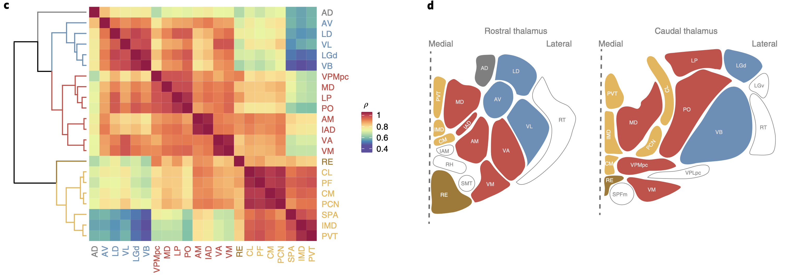
Figure 2: Map of thalamic areas in the rodent according to gene expression patterns, which identifies 3+1 major clusters. Blue are primary sensory and other areas that receive strong subcortical driver inputs. Red are higher-order areas that either receive cortical drivers or no drivers. Orange are the intralaminar and paraventricular nuclei that have more specialized connectivity with the basal ganglia and other subcortical areas. The reunions nucleus, which interconnects the prefrontal cortex with the hippocampus, has its own unique status. From Phillips et al., 2019.
Figure 2 shows a map of different thalamic areas in rodents, which provides a broad set of 3+1 categories based on patterns of gene expression, which is useful for informing our understanding of function across different thalamic areas. This data does not provide more fine-grained distinctions in terms of patterns of connectivity, but they do generally correspond to different types of driver inputs, as we’ll see below.
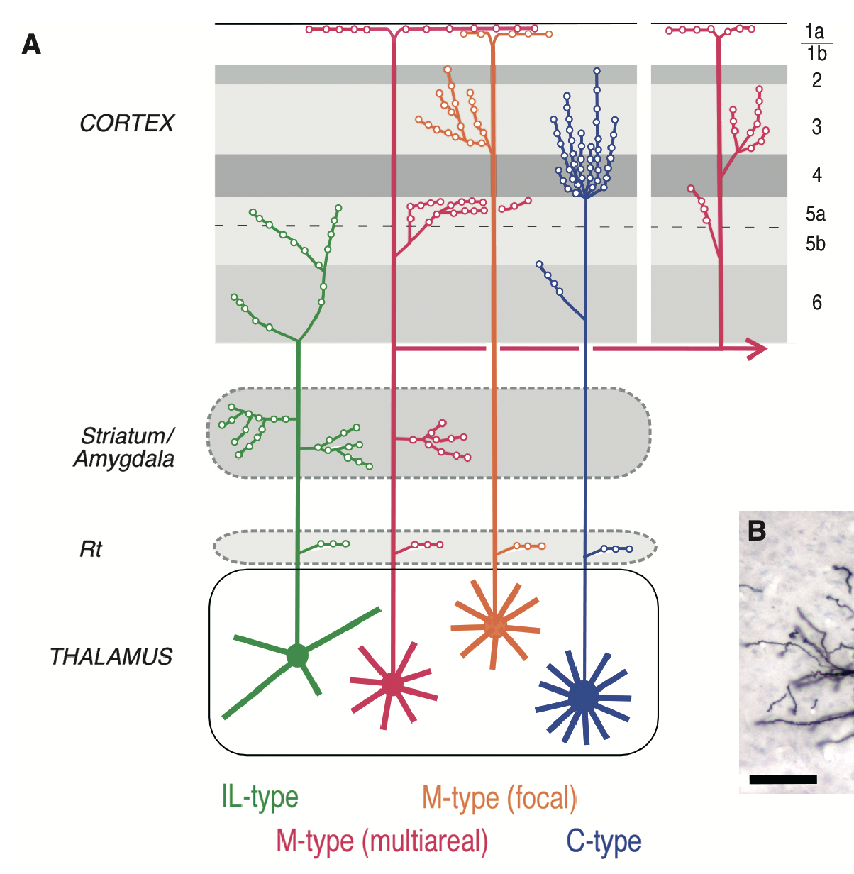
Figure 3: Different types of thalamic projections into the cortex, varying in the laminar targets and the area focus, according to Clasca et al., 2012. The C-type is the classical first-order sensory core-type, projecting to the middle lamina in a focal manner, while the M-type (multiareal) is the classical matrix type projecting to layer 1 and 5, across multiple areas. The M-type (focal) combines features from classical core and matrix types, with projections that target middle layers in addition to layer 1, with more focal area-wise connectivity. Meanwhile the IL (intralaminar) pattern (i.e., PF = parafascicular thalamus) is specific to neurons in these nuclei, targeting only deep layers and also extensively the striatum.
Figure 3 shows an extension of the classical core and matrix categories by Clascá et al. (2012), with two distinct M-types, one of which corresponds to the classical matrix type, while the M-type (focal) combines features of core and matrix. There is also the unique projection pattern of the intralaminar (IL) nuclei, which includes the PF (parafasicular) and CM (centromedian) nuclei, which play a critical role in the function of the basal ganglia (see Parafascicular feedback loops), and have extensive connectivity into the striatum along with projections into the deep cortical layers.
Instead of retaining a core-vs-matrix categorical approach, we prefer to use functional, feature-based terminology in describing the connectivity profiles of neurons in different thalamic areas. A given TRN cell will in general have multiple of these properties.
• Input: focal projections into the middle layers, driving informational input to a cortical areas.
• Maintenance: focal projections to layers 1 and 5, supporting active maintenance in layer 5b PT neurons.
• Modulatory: diffuse projections to layers 1 and 5. This is not exclusive to classical arousal-modulating matrix-type neurons, as basal-ganglia modulated motor areas including VA and VM have this pattern, to provide more dynamic, fine-grained modulation of motor cortical areas.
• Single: projects to a single area.
• Multi: projects to multiple areas. Note that this can be orthogonal to the focal vs. diffuse nature of the projection, for example MDm (mediodorsal medial) has focal but multi-area projections.
Functional organization of the thalamus
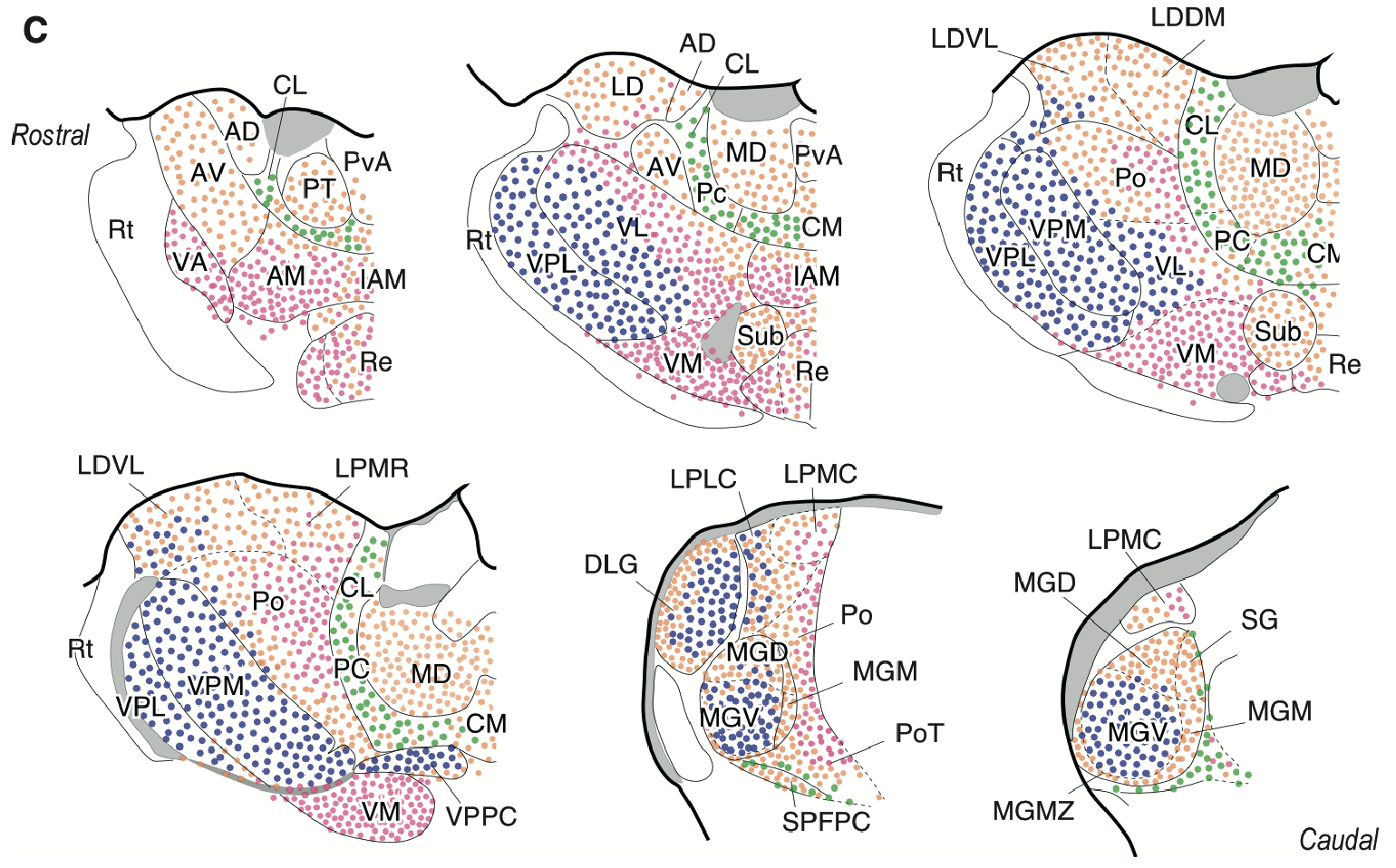
Figure 4: Map of thalamic areas in the rodent according to the four types shown in the prior figure, using the same colors. From Clasca et al., 2012.
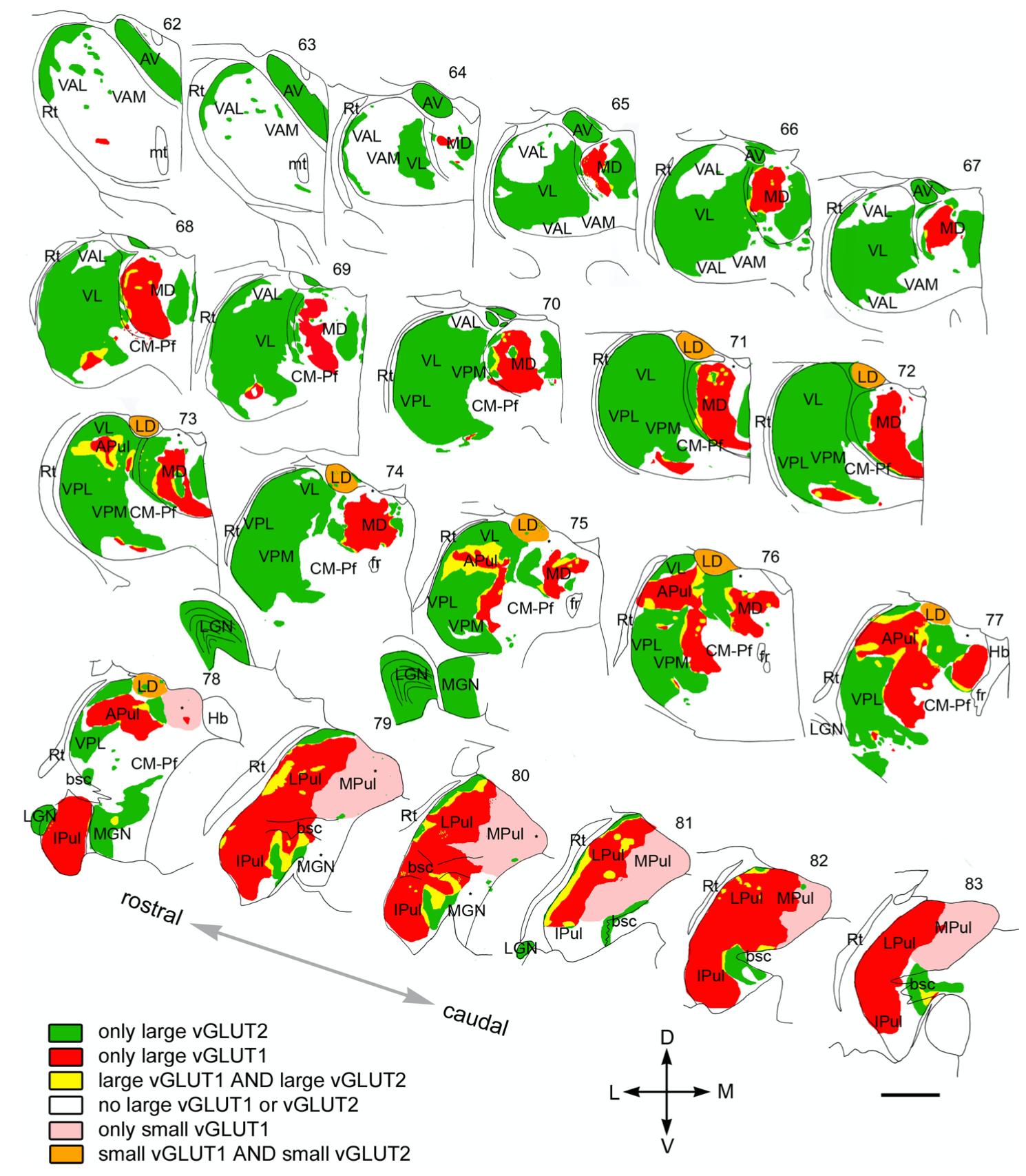
Figure 5: Different types of thalamic drivers in the primate thalamus, which provides key insight into functions of these thalamic areas and the cortical areas they interconnected with. Large = strong, small = weak. vGLUT2 = subcortical drivers, e.g., green are all primary sensory (LGN = vision, MGN = auditory, VPM, VPL = somatosensory) and motor (VL = cerebellum); vGLUT1 = cortical drivers (red). Inhibitory driver inputs, as from the basal ganglia output areas (SNr, GPi, VP), show up as blank (white) (e.g., the VAL, VAM, VAL frontal thalamic areas), and the MD actually has a mix of neurons with these inhibitory drivers and pulvinar-like cortical drivers, but only the pulvinar-like aspect is represented as indicated by its red color, except the medial MDm which has more uniform inhibitory input from VP (ventral pallidum). From Rovo et al., 2012.
Figure 4 provides a map of the rodent thalamus using the Clascá et al. (2012) categories, and Figure 5 provides a map of the macaque monkey thalamus, labeling the type of driver synapse present in each area (Rovó et al., 2012). These are functionally interpretable as follows:
- • Subcortical drivers: Strong = large vGLUT2 (green).
- • Cortical drivers: Strong = large vGLUT1 (red), Weaker = small vGLUT1 (pink).
- • None (white). These typically receive inhibitory inputs from the basal ganglia.
- • Other combinations are discussed below where applicable.
By putting these two maps together (and keeping in mind the data in Figure 2), we can identify the general functionality of different thalamic areas.
It is critical to recognize that although each thalamic nucleus has a predominant type of TRN connectivity pattern that we identify it with, there is almost always some diversity, such that for example classical matrix-type neurons are intermixed within the classical core-type that predominate in the LGN (Sherman & Usrey, 2024). Functionally, these matrix-type neurons can cover a lot of territory with their broad connectivity patterns, so fewer are required to do the job. In general, we assume that modulation of overall excitability and arousal levels in the cortex via such neurons is an additional job performed by most thalamic areas, but dedicated areas that perform only this function are unnecessary.
• Primary sensory (Driver: Subcortical; Projection: Input, Single): LGN (lateral geniculate nucleus, vision, V1); MGNv (medial ventral GN; auditory, A1); MGNm (medial medial GN; vestibular and others); VPL, VPM (ventral posteriolateral, lateral, medial; somatosensory, S1). These are most of the green areas in the driver figure, with the exception of VL and AV, having large, strong glutamatergic drivers from subcortical sources (e.g., the retina for LGN).
• Primary motor (Driver: Subcortical; Projection: Input, Single): VL (ventral lateral). This is a primary, first-order type of thalamic area, which instead of being sensory, is primary motor (M1). The strong excitatory subcortical drivers onto these neurons arise from ascending spinal pathways that converge onto the cerebellar nuclei (CN) and then project from there into the VL. These neurons provide multiple training signals to primary motor cortex, including efferent copy signals reflecting what the spinal motor system actually did, versus the commands that were sent down to it. Anatomically, VL is adjacent to the somatosensory VPL, VPM nuclei which project topographically to primary somatosensory cortex (S1), just across the central sulcus from M1. See below for further discussion of the frontal thalamus areas.
• Primary space and time (Driver: Subcortical; Projection: Input, Multiple): AV (anteroventral) and AD (anterodorsal). Like the VL, this is an interesting case of a subcortically driven, input-projecting area that is not a standard sensory input. These areas receive driver inputs from the mammillary bodies (medial to AV and lateral to AD), which convey important spatial and timing signals, that are then projected into the (pre-post) subiculum and retrosplenial cortex, which are the primary spatial representation areas of the cortex. The lateral mammillary bodies projecting to the AD nucleus are the primary source of head direction cells, that integrate vestibular and motor signals to keep track of which way the head (and body more generally) is facing in an allocentric (world-based) coordinate system. The medial mammillary bodies projecting to the AV convey the theta rhythm signals generated in the ventral tegmental nucleus (VTN), which also drive neurons in the medial septum, to coordinate processing throughout the medial temporal lobe and hippocampus.
• Secondary perceptual (Driver: Cortical; Projection: Input, Modulatory, Single & Multi): Pulvinar is the main nucleus of this type, with topographic projections into all of the higher-order sensory areas. The driver map shows these areas in red and pink, where the red areas have strong glutamatergic drivers, but unlike primary areas, these come from the layer 5b cortical drivers, not subcortical drivers. The pulvinar as such is not labeled as such in the rodent, but the lateral posterior (LP) is thought to be a homologous area based on connectivity (Leow et al., 2022), and it has the same focal connectivity (LPMC, LPMR). The yellow areas in the driver map represents areas of pulvinar that receive both cortical and subcortical inputs, specifically from the superior colliculus, which is known to project into the pulvinar.
• Secondary motor (Driver: None; Projection: Modulatory, Maintenance, Input, Multi & Single): VA / VM, AM (ventral anterior, medial and anterior medial, including VAM, VAL Figure 5) are all colored white in the driver figure, because they have no strong drivers. Instead, they receive convergent input from a range of different subcortical sources, along with extensive inhibition from the basal ganglia output nuclei (SNr, GPi). These are all of the red dot areas in the connectivity figure, representing the main population of M-type multiareal neurons. Based on this pattern of connectivity, these are likely to have very broad modulatory effects on the corresponding higher-order motor areas, allowing the BG to modulate overall excitability in these motor cortex areas via disinhibitory gating of the BG output pathways. See frontal thalamus below for further discussion.
• Secondary PFC, goal-gating (Driver: None; Projection: Input, Maintenance, Modulatory, Multi): MD (mediodorsal). In rodents, different areas of MD (medial, central, lateral) receive different BG disinhibitory gating signals, and project in a multi-area yet focal manner to the middle lamina of all of the goal-driven areas of PFC (Kuramoto et al., 2017; Phillips et al., 2021), supporting multiple levels of goal-gating. Area MDm receives extensive projections from the ventral pallidum (VP) (showing up as white in Figure 5) and projects to all goal-driven PFC areas, representing the primary goal gating area. The MDc and MDl (and associated areas in primates) have more varied connectivity and are discussed next.
• Secondary PFC, predictive learning and goal updating (Driver: Cortical; Projection: Input, Maintenance, Single): MDc, MDl (mediodorsal central, lateral). A subset of neurons within these areas of MD are effectively like the pulvinar for prefrontal cortex, in terms of receiving strong cortical driver inputs (red in Figure 5), while having M-type focal connectivity patterns. Based on the strong theoretical and empirical basis for pulvinar supporting predictive learning, this same function is likely supported for the goal-driven areas of OFC, IL, PL, and ACC PFC that these MD areas project to. The other neurons in these MD areas receive extensive disinhibitory BG input from GPi and SNr, and each project to the two separate components of the goal-value system: OFC, IL = outcome value (MDc); ACC, PL, ALM / dlPFC = action value / cost (MDl).
• Intralaminar (IL: PF, CM, etc) (Driver: None; Projection: IL-type to striatum, cortical deep layers). These nuclei provide extensive feedback to the basal ganglia and also to deep layers of the motor neocortex, and play critical specialized roles in BG function and learning.
Giguere & Goldman‐Rakic, 1988 is primate study showing patchy organization in primates.
Frontal thalamus
There are three major functions for thalamic areas interconnected with frontal areas:
• Training of primary motor cortex to speak the language of the spinal cord (VL, POm / PULm). These receive strong driver input from spinal motor signals (VL) or somatosensory signals (POm / PULm), and project strong driver inputs into the middle lamina of motor areas.
• Corticocortical predictive learning, analogous to that supported by the pulvinar for posterior cortex, so higher layers can learn to predict the motor-related activity of lower layers. This is supported by subsets of neurons within the MD and AM nuclei. This helps learning of longer sequences of motor actions, for example.
• Two forms of basal ganglia (BG) based rapid updating (gating) of frontal representations across multiple timescales: activation-based working memory updating for goal engagement (per the Rubicon model) and motor plan updates, consistent with the PBWM (prefrontal cortex, basal ganglia working memory) model; and a more diffuse, broad synchronization form of gating, associated with motor action initiation or plan updates. The working memory gating function is supported by subsets of neurons within MD and AM that receive BG disinhibitory outputs, and the broad synchronization function is supported by the VA (aka VM in rodent; Ilinsky & Kultas‐Ilinsky, 1987) nucleus, including the special VAmc nucleus that is specific to primates.
Rodent
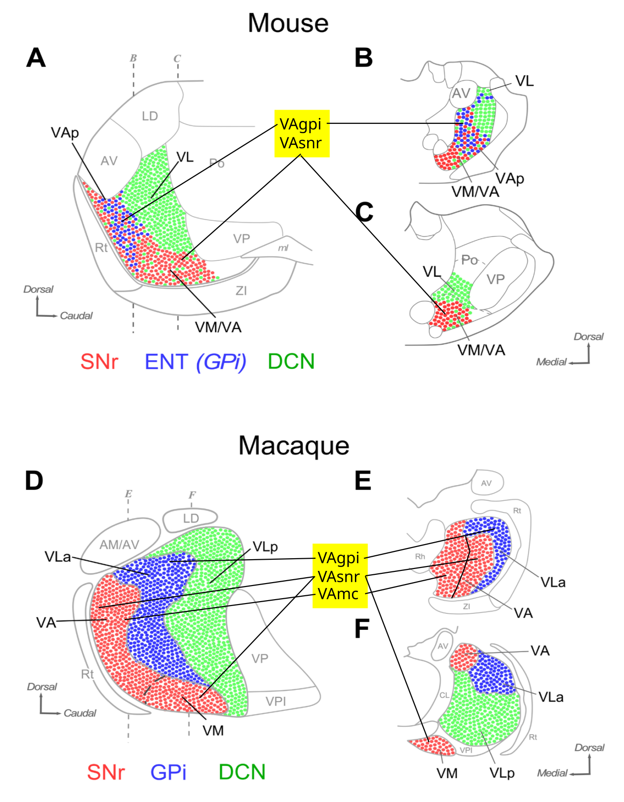
Figure 6: Summary of inputs to the ventral anterior thalamic nuclei that interconnect with the frontal cortex, in mouse (rodent) and macaque (primate). A and D are saggital plane, while the others are coronal slices. The highlighted labels in the middle provide an alternative to the inconsistently used anatomical labels, based definitionally on the source of the driver inputs: VAgpi is the part of VM/VA that receives from GPi, while VAsnr receives from SNr. The VL in rodent can be clearly defined as the area receiving strong driver inputs from DCN (deep cerebellar nuclei). The macaque data is schematic from older sources. VAmc is the medial portion of VA as shown, which only exists in primate, and is defined by its projections into the PFC, whereas VAgpi and VAsnr correspond with the rodent versions, which only project to motor areas. VLp is the DCN recipient area, which can be labeled VLcn for clarity. From Alonso-Martinez et al., 2023.
We begin with the simpler case of the rodent, which has each of these different functions clearly represented by different thalamic nuclei. The primate case then adds more complexity that can be understood as building upon this same basic foundation. Figure 6 shows the organization of BG (SNr, GPi) vs. cerebellar (CN) driver inputs for the ventral anterior thalamic nuclei that project to the frontal cortex (Alonso-Martínez et al., 2023). Because the anatomical terminology for these thalamic areas is inconsistent across papers and across species, we define the following labels that will be used in the remainder of our discussion (see Sieveritz & Kiani, 2024 for a similar organization):
- •
VAbgare VM/VA/VL areas that receive inhibitory inputs from BG output nuclei, and project to motor cortex but not PFC areas, specifically: - ◦
VAgpireceives inhibitory inputs from GPi (entopeduncular or ENT in rodent). - ◦
VAsnrreceives inhibitory inputs from SNr. In rodents, this includes rostroventral VA (Alonso-Martínez et al., 2023). - •
VAmcis the subset of VA in primate that has magnocellular cells, which projects to PFC and receives from SNr (does not exist in rodent). - •
VLcnis the VL area that receives excitatory driver inputs from CN (cerebellar nuclei), and projects to primary motor (M1). In rodents, this also includes caudodorsal VA (Alonso-Martínez et al., 2023).
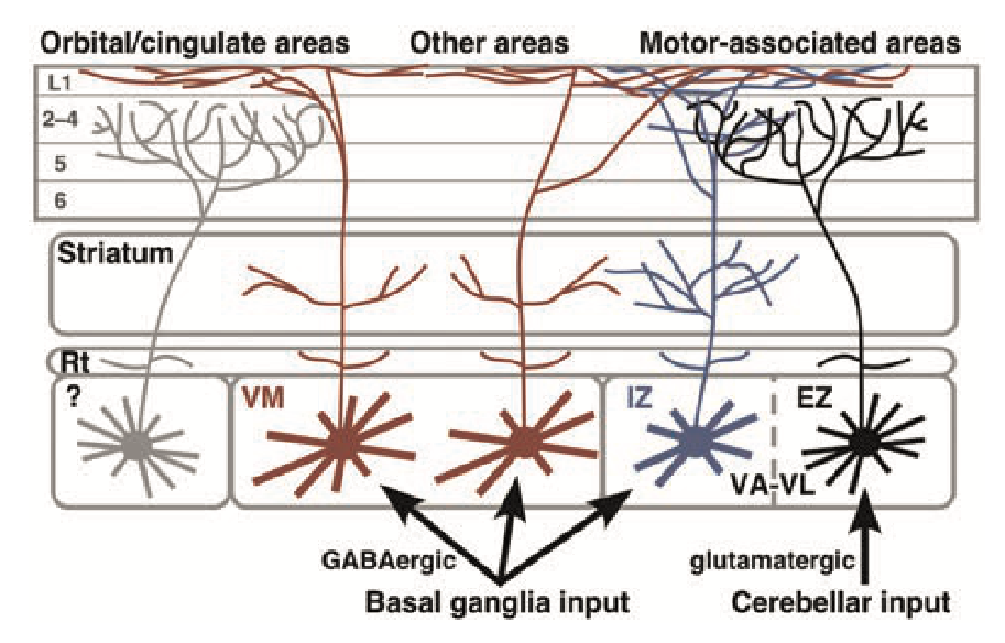
Figure 7: Summary of projections from the different ventral motor thalamic areas in rodents. VL in the EZ = excitatory zone is thus the VLcn that receives cerebellar nucleus (CN) driver inputs (which includes caudodorsal VA in rodent), and sends focal core-like projections as shown on the right. The VM neurons (which include rostroventral VA, in the IZ = inhibitory zone) represents VAbg (VAsnr, VAgpi) in our terminology, because it receives extensive BG disinhibitory output (SNr, GPi) and projects in a more diffuse matrix-like pattern to superficial layers. From Kuramoto et al. (2015).
Figure 7 (from Kuramoto et al., 2015) shows ventral thalamic pathways in rodents, which can be identified according to their driver inputs, per our standard labels abovel. VLcn receives strong drivers from the cerebellar nuclei, and projects focally to middle lamina in motor areas (i.e., core-like), enabling motor areas to learn the language of the spinal cord. This projection goes to M2, SMA and premotor areas in the primate and corresponding higher motor areas in the rodent (Ilinsky & Kultas‐Ilinsky, 1987).
VAbg receive BG disinhibitory outputs (VAsnr = SNr, VAgpi = GPi), and project to superficial lamina in a widely branching manner (i.e., matrix-like), which allows the BG to send a broad gating signal to all of motor cortex, and even beyond, with some neurons projecting to widespread areas in posterior cortex as well. Furthermore, there was some indication of a subset of these neurons, within the inhibitory zone of VA-VL, that had more restricted connectivity favoring OFC and ACC areas (Kuramoto et al., 2015). See the next section for more discussion, but overall these neurons appear to be important for synchronizing all of the motor areas at the point of important transitions, such the transition between motor preparation to motor execution. See
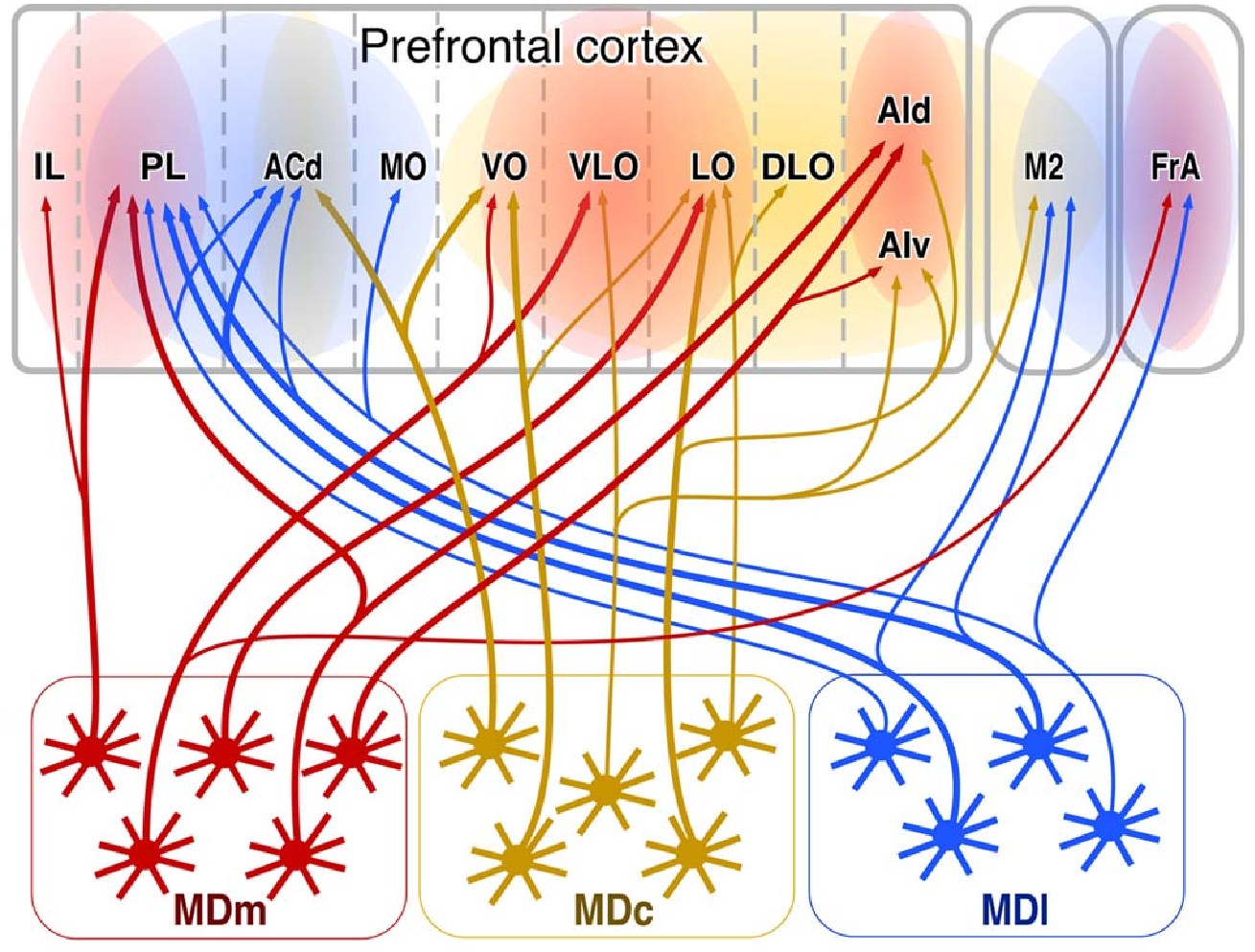
Figure 8: Thalamocortical connectivity for different regions of the MD thalamus in rats. MDm (medial) is the main goal-driven gating area, which receives disinhibitory input from ventral pallidum (VP), and projects to the core goal areas (IL, PL, and OFC areas). MDc (central) projects to more OFC areas and may be important for updating during goal approach. MDl (lateral) targets the complementary ACC and PL goal areas, along with motor plan areas in (FrA = frontal association higher level motor, M2 = secondary motor). From Kuramoto et al., 2017.
Figure 8 shows the different regions of the MD thalamus interconnecting with areas of the PFC in rats (from Kuramoto et al., 2017), with the primary goal-gating functionality supported by MDm (medial), which projects to all of the goal areas (IL, PL, OFC, and FrA (ALM)) and receives disinhibition from VP (ventral pallidum), which is the output pathway from ventral striatum goal gating areas as described in basal ganglia. The other two areas of MD (c = central, l = lateral) provide complementary, more specific gating pathways for OFC (central) and motor, ACC, and PL (lateral), potentially allowing each of these coordinated components of the goal representation to be updated separately. The MD projections are much stronger onto middle layers (2,3) compared to projections from the VA (VM), which target layer 1 and deep layers (Collins et al., 2018).
These MD areas also have non BG-gated neurons that putatively receive CT inputs (from layer 6) and cortical drivers (from layer 5 PT neurons), and function just like the pulvinar for predictive learning (Collins et al., 2018). The Rubicon model organizes these so that e.g., more abstract IL value neurons predict OFC specific outcome representations, while PL predicts both IL and ACC activity. ACC learns by predicting subcortical action cost signals, while OFC learns by predicting BLA (amygdala) outcome signals.
The posteriormedial (POm) nucleus in the rodent is a higher-order thalamic area for somatosensory signals from primary sensory cortex (S1), similar in connectivity to the pulvinar in relation to visual and other higher-order sensory processing areas (Mo et al., 2017; Landisman & Connors, 2007). For example, only POm receives driver inputs from layer 5b PT neurons, compared to the primary somatosensory thalamic nucleus (VPM).
Primate
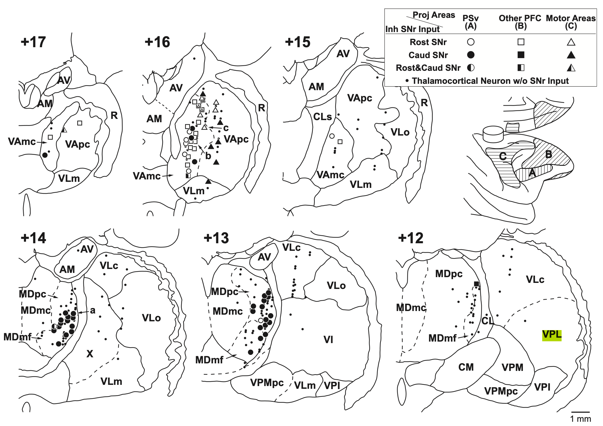
Figure 9: Thalamic nuclei in macaque monkeys showing the projections received from SNr (BG) as a function of the frontal areas that these cortical areas project to (inset below legend), with A = PSv, ventral of the principal sulcus (e.g., IFG in human PFC); B = dlPFC (areas 9, 46); C = motor areas (M1, SMA, PMd). VAmc projects widely to all PFC areas, and has significant SNr input. VApc corresponds to VAsnr in our terminology, and projects only to motor areas. MDpc and MDmf mainly interconect with the PSv area associated with caudal SNr inputs. AM = anteromedial. Note that MD has a mix of neurons with no SNr input (typically with cortical drivers, like pulvinar), and those with SNr or VP (ventral pallidum, for MDmc) inputs. From Tanibuchi et al., 2009.
Figure 9 (from Tanibuchi et al., 2009) the anatomical organization of the frontal thalamic areas in the macaque monkey, including the different subdivisions of the MD (where the rodent MDm = MDmc, MDc = MDpc, and MDl = MDmf, MDdc), VLcn (including VLm and VLo, defined functionally as neurons receiving driver inputs from cerebellar nuclei), and the primate-specific VAmc (medial, magnocellular portion of VA) that projects to the PFC, and rodent-like VAgpi, VAsnr in our terminology, which project only to motor areas, and maps onto VApc and portions of LVo in this primate terminology.
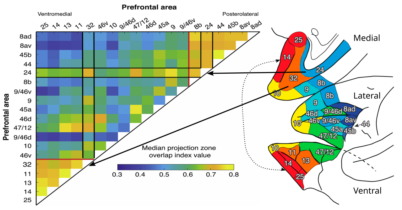
Figure 10: Similarity of patterns of MD thalamic connectivity with prefrontal cortex (PFC) in the macaque (which typically also reflects direct interconnectivity strength), which clearly delineates the premotor, dlPFC & vlPFC, and the goal-driven areas. Red and orange areas are ventral and medial PFC goal-driven areas (i.e, the core of the Rubicon model), including OFC (13, 14, 11; specific outcomes), IL (25; abstract outcomes), and PL (32; integrated reward-cost utility: note broader interconnectivity, especially with 24, 25 and 9); Yellow (10) is polar frontal cortex, unique to primates, important for episodic memory and outer loops of processing; Green (47,12) is vlPFC (ventrolateral) for control over the temporal lobe (language, objects etc); Cyan (9, 46d, 45a) is dlPFC (dorsolateral) for control over parietal lobe for spatial and temporal organization of action (broad plans); Blue (8) is FEF (frontal eye fields) which has similar connectivity to PM (premotor), SMA (supplementary motor area) and primary motor cortex (M1). Area 24 is ACC (anterior cingulate cortex) which represents the value (mostly costs) of actions, and has very broad interconnectivity with motor areas, but also goal areas, especially 32 (PL). Adapted from Phillips et al., 2019.
Figure 10 shows the similarity structure of the various PFC areas according to their patterns of MD thalamic connectivity (Phillips et al., 2019). This identifies the primary functional groupings of goal-driven areas in v/mPFC (OFC, IL, PL), the ACC which bridges motor and these goal-driven areas to represent action value (costs), and the dlPFC and vlPFC that interconnect with posterior cortical areas (parietal and temporal lobes, respectively; O’Reilly, 2010) and guide motor frontal actions (premotor areas shown).
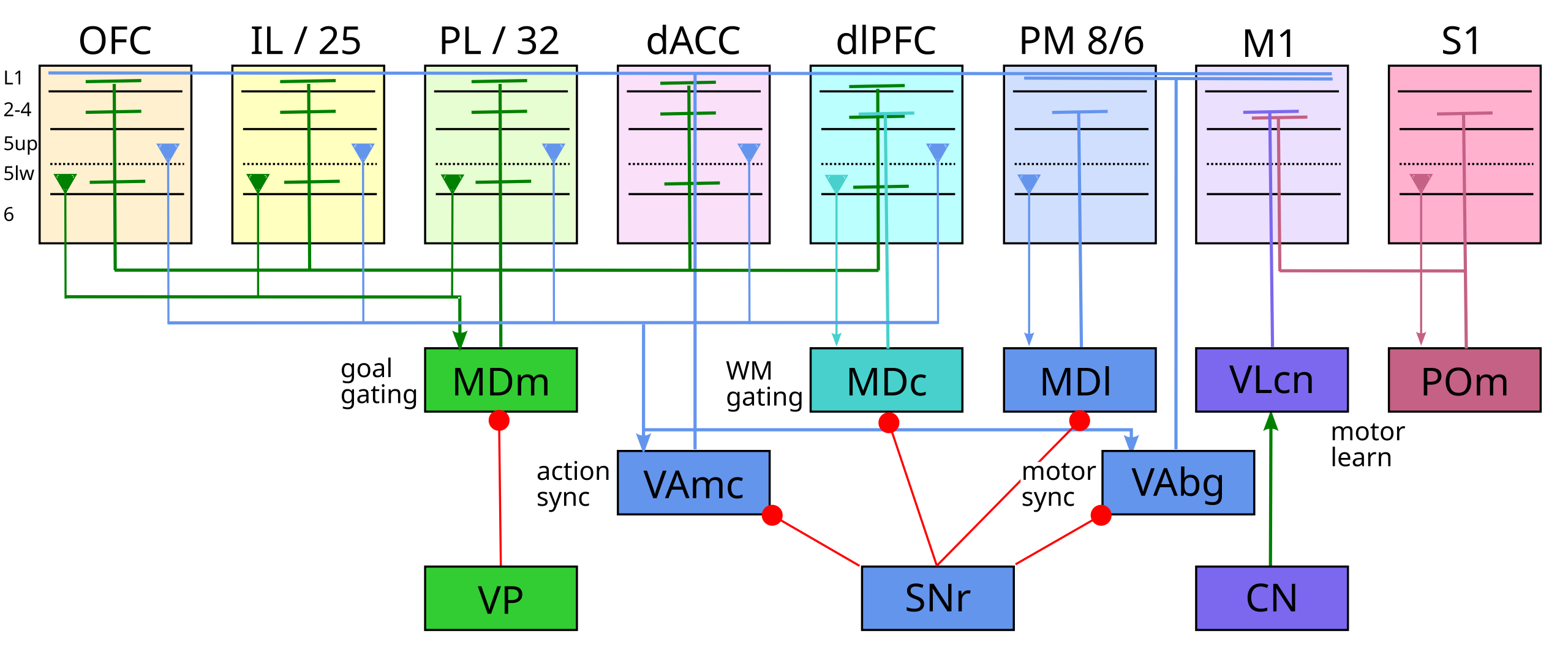
Figure 11: Summary of frontal thalamic connectivity and function for the primate case (macaque). MDm (medial) is the goal gating nucleus, receiving from and projecting to all the goal-driven PFC areas (OFC, IL, PL, dACC, dlPFC), with disinhibitory gating from the ventral pallidum (VP). MDc (central) drives dlPFC working memory (WM) gating (and, separately, OFC updating as in the rodent, not shown), via SNr disinhibition. MDl does premotor (PM, SMA areas 8,6) gating (and separately PL, ACC as in the rodent, not shown). MD cortical drivers originate in layer 5b lower (5lw). VLcn has cerebellar drivers and projects to M1 to train motor representations to speak the language of the spinal muscle synergies. POm (posterior medial in rodents, medial pulvinar in primates) drives predictive learning in motor cortex from somatosensory (S1) drivers. VAmc and VAbg (VAsnr, VAgpi) have broad convergent inputs from respective frontal areas, originating from the upper layer 5b PT neurons (5up), and project matrix-style modulator outputs to layer 1 across a broad range of areas (PFC for VAmc, motor areas for VAbg). This is likely important for synchronizing these areas at major points of transition, e.g., between planning and execution of actions.
Table 1: Frontal thalamus areas
| Area | Function | Drivers | Modulators | BG | Strong outs | Weak outs | Lamina | Other ins | Other outs |
|---|---|---|---|---|---|---|---|---|---|
| Learning | |||||||||
| PULm / POm | S1->M1 Learn | S1, Spinal | M1, M2 | – | M1, M2, SMA.. | Mid | spinal somatosensory | ||
| VLcn | CN->M1,M2 Learn | CN | M1,M2 | – | M1, M2 | Mid | CN | ||
| MD | Predictive learning | (see below) | – | ||||||
| Gating | (focal) | ||||||||
| AM | MTL <-> goal | ACC, OFC, IL, PL | d/vlPFC, lOFC | mGPi | 10, 11, 12, 46 | PL, ACC | Mid | CA3, aSub, BLA, mGPi | vSub, RSC |
| MDm (mc) | goal engage | OFC, IL, PL | vlPFC, ACC | VP | OFC | PL, IL | S/D, Mid | IT polar, EC, PRh, Sub, BLA | |
| MDc (pc) | dl/plan WM | 10, dlPFC | ACC, PL, PM, SMA | SNr | dlPFC (WM) | PM, SMA | Mid | parietal | parietal |
| MDl (mf,dc) | eye/act motor | 8 (FEF), PM, SMA | SNr | 8, PM, SMA | Mid | SC, parietal attn | |||
| Gating | (diffuse) | ||||||||
| VAgpi, VAsnr | BG->motor | GPi, SNr | M1, M2, SMA, DM | ||||||
| VAmc | BG->dl/motor | dl, PL, ACC | SNr | dlPFC, M2, M1 | Mid + S/D | parietal, temp pole | parietal |
Figure 11 and Table 1 provide an overall summary of all motor thalamus areas in primate and rodent, organized according to the three primary functions listed above, based on all the available data (Phillips et al., 2021; Timbie & Barbas, 2015; Xiao et al., 2009). Note the dual-functionality of area MD, for pulvinar-like predictive learning and BG-driven gating / updating, which is consistent with 80% of the projections to MD arising from layer 6 CT neurons, and 20% from deep layer 5b (PT bursting neurons) (Xiao et al., 2009).
The function of the MDc in primates has been expanded relative to the rodent case (where it primarily targets OFC), to include a significant role in updating working memory representations in the dorsal and ventral lateral areas (dlPFC, vlPFC) (Funahashi et al., 1989; Fuster & Alexander, 1973; Takeda & Funahashi, 2002), consistent with the PBWM (prefrontal cortex, basal ganglia working memory) model. It is likely that different neurons target different areas within the large expanse of MDc, so the rodent-specific case is likely still supported by a subset of these neurons. Likewise, MDl in primates includes a more significant role for saccade-based updates of motor representations, driven by strong FEF 8a driver inputs, which could then drive broader motor gating.
The other major new element in the primate thalamus is the VAmc, which is the magnocellular compartment of the ventral anterior thalamus (Phillips et al., 2021). This area has very broad overlapping inputs, with virtually no topographic organization of input projections, which come from all over the PFC, and project back broadly to the PFC and to motor cortex. Consistent with this broad diffuse connectivity, it sends matrix-like layer 1 outputs, but also projections to middle lamina.
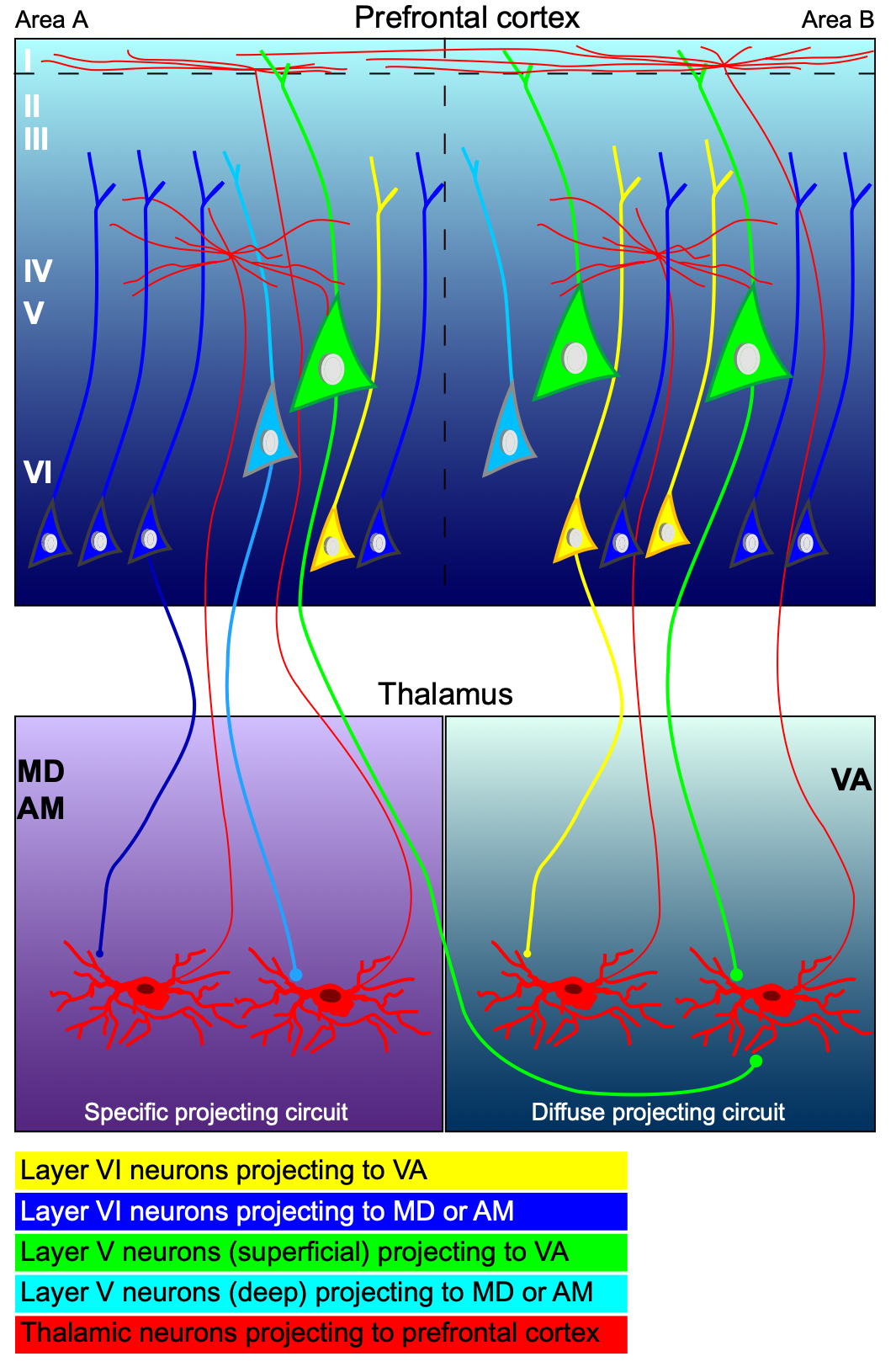
Figure 12: Different patterns of projections to and from thalamic areas MD & AM vs. VA. MD and AM project more focally, into both the middle cortical lamina and the superficial, with different thalamic neurons having different preferred targets (see also Mukherjee et al., 2021), whereas VA projects diffusely, also into both target lamina. Critically, the layer 5 (V) neurons that project into MD & AM originate in the deep half of 5b (light blue), while those that project into VA originate in the superficial half (green). Economo et al. (2018) showed that these superficial neurons are more likely to be active during preparatory periods, while the deep half also project subcortically (i.e., to motor targets) and are preferentially active during execution. The layer 6 (VI) neurons preferentially project into the middle-lamina projecting thalamic cells, although in MD & VA these also receive cortical drivers from other areas (not shown). From Xiao et al. (2009).
Interestingly, the layer 6 CT inputs to VAmc target the PV expressing neurons that project to the middle layers of cortex (core-style), while the layer 5 inputs arise from layer 5b upper PT neurons (Xiao et al., 2009; Economo et al., 2018; Hattox & Nelson, 2007; Figure 12) that target CB expressing neurons, which give rise to the layer 1 superficial and some deep layer projections (matrix-style). As discussed in Economo et al. (2018), this subtype of 5b PT neurons is generally more active during motor preparation and planning, while the layer 5b lower PT neurons are more active during motor execution, and correspondingly send projections to the brainstem. These lower subtype are the neurons that project to the MD and AM thalamus (Xiao et al., 2009), consistent with the idea that the VA (VAmc, VAgpi, and VAsnr) areas are functionally distinct from MD and AM, as shown in Figure 12.
The broad connectivity of VAmc, similar to that of VAbg, seems ideally suited for synchronizing processing across the frontal motor and PFC areas according to major motor-based events, such as saccades or large-scale body movements. There are generally abrupt transitions in cortical states at the onset of motor actions, and these broad, diffuse connections could effectively broadcast signals that facilitate these transitions, enabling the system to transition between preparatory activity and actual motor action (Churchland & Shenoy, 2024; Economo et al., 2018). A growing literature on meta-stable neural activity states with rapid transitions between relatively stable intervening states is consistent with this dynamic (La Camera et al., 2019; Recanatesi et al., 2022).
The role of the AM in interconnecting the medial temporal lobe including the hippocampus, subiculum, and the retrosplenial cortex (RSC) in the medial parietal lobe is critical for enabling episodic memories to be engaged in the service of goal planning, as is discussed a bit further in the section on anterior thalamic areas (see also Xiao et al., 2009).
Liu et al., 2024 show that OFC -> V1 hits PT lower output neurons, projecting subcortically, while LP (Pulvinar) -> V1 hits PF upper IT neurons.
Anterior thalamic areas
Aggleton & O’Mara, 2022 – key ref.
- • AM: anteromedial, is the bridge between MTL and goal areas in PFC, see Table 1
Burst firing
Thalamic relay cells have slow T-type calcium channels that can produce burst firing after periods of hyperpolarization (Llinas & Jahnsen, 1982; Destexhe et al., 1998), which inspired a hypothesis from Francis Crick that these bursts are critical for attentional modulation (i.e., as a kind of “searchlight”) in cortex (Crick, 1984). However, these T-type calcium channels require a period of hyperpolarization to activate them, after which the burst arises as a rebound firing phenomenon. This hyperpolarization is typically present during sleep, not during wakefulness, and bursting is far more prevalent during sleep and inactive states.
Thus, a simple account is that bursting is only present during sleep or similar inactive states, as a way of driving strong synchronous activity waves (“slow waves”) that characterize most phases of sleep. This synchronous activity largely prevents information processing by disrupting the ability to activate sparse distributed representations with stable activation states, which are essential for meaningful computation.
Nevertheless, relatively low rates of thalamic bursting do occur in the awake, behaving state (Guido & Weyand, 1995; Ramcharan et al., 2005; Boshra et al., 2025), raising the question as to whether they may be functionally relevant (e.g., Zeldenrust et al., 2018). One hypothesis is that they may provide a kind of “wake up” effect, exciting the cortex more when it previously was inactive or new information is activating different thalamic neurons (e.g., Sherman, 2001; Sherman, 2001). However, Steriade (2000) observed that:
If the “wake-up” call hypothesis were functioning, the bursts should wake us more efficiently as we progressively fall into deeper stages of sleep. This would indeed be inconvenient.
The counter-point here is that the broad levels of synchrony in this bursting and overall population activity are really the critical drivers of sleep-state processing, and that bursting during awake behavior is much sparser and more distributed in time and across neurons, and therefore would not drive these levels of synchronous firing.
From studies that manipulated burst activity and measured the effects on cortical targets, it is clear that bursting does indeed have the kind of excitatory impact on cortex that one might otherwise expect from a rapid sequence of excitatory inputs (Alitto et al., 2019; Swadlow & Gusev, 2001; Boshra et al., 2025).
Therefore, putting together the properties of the T-type calcium channels and this greater downstream impact of bursting excitation, it does make sense that there would be some amount of augmented attention from bursting driven by novel inputs. However, because of the hyperpolarization requirement for activating the T-type channels, anytime there is any kind of sustained ongoing activity, bursting should not be a major contributor.
The importance of baseline activity level in determining burst probability is illustrated by studies that compared primary sensory thalamus (which will generally have a high level of sensory-driven activity) versus higher-order thalamic areas, which may be more sparsely activated depending on what kind of task is being performed. One early report (Guido & Weyand, 1995) showed a burst probability of just under 1% in the primary visual cortex of awake, behaving cats. Consistent with the above account, they also report that most of these occurred at the onset of visual stimulation.
Ramcharan et al. (2005) reported higher levels of bursting (10-20%) for neurons in higher-order thalamus (pulvinar and MD) than first-order sensory thalamus (LGN, VP, MGNv), in awake but barely behaving monkeys (head fixed, either sitting idle or holding a simple fixation). Boshra et al. (2025) showed that bursting in the pulvinar in awake behaving monkeys was more likely for idle periods, but showed a small increase for attended locations. Although they emphasized the effects of bursting driven by electrical stimulation, it was not clear that purely naturalistic bursting had clear behavioral implications.
In conclusion, it seems likely that thalamic bursting is mostly relevant for sleep, and thus can be safely omitted from models, incurring a small penalty as an error of omission. The behavioral effects are likely to be relatively modest, because new activity from previously inactive thalamic neurons will likely already be more impactful in driving cortical neurons.